- You are here: Home
- Resources
- Explore & Learn
- Cell Biology
- Monocytes vs. Macrophages
Support
-
Cell Services
- Cell Line Authentication
- Cell Surface Marker Validation Service
-
Cell Line Testing and Assays
- Toxicology Assay
- Drug-Resistant Cell Models
- Cell Viability Assays
- Cell Proliferation Assays
- Cell Migration Assays
- Soft Agar Colony Formation Assay Service
- SRB Assay
- Cell Apoptosis Assays
- Cell Cycle Assays
- Cell Angiogenesis Assays
- DNA/RNA Extraction
- Custom Cell & Tissue Lysate Service
- Cellular Phosphorylation Assays
- Stability Testing
- Sterility Testing
- Endotoxin Detection and Removal
- Phagocytosis Assays
- Cell-Based Screening and Profiling Services
- 3D-Based Services
- Custom Cell Services
- Cell-based LNP Evaluation
-
Stem Cell Research
- iPSC Generation
- iPSC Characterization
-
iPSC Differentiation
- Neural Stem Cells Differentiation Service from iPSC
- Astrocyte Differentiation Service from iPSC
- Retinal Pigment Epithelium (RPE) Differentiation Service from iPSC
- Cardiomyocyte Differentiation Service from iPSC
- T Cell, NK Cell Differentiation Service from iPSC
- Hepatocyte Differentiation Service from iPSC
- Beta Cell Differentiation Service from iPSC
- Brain Organoid Differentiation Service from iPSC
- Cardiac Organoid Differentiation Service from iPSC
- Kidney Organoid Differentiation Service from iPSC
- GABAnergic Neuron Differentiation Service from iPSC
- Undifferentiated iPSC Detection
- iPSC Gene Editing
- iPSC Expanding Service
- MSC Services
- Stem Cell Assay Development and Screening
- Cell Immortalization
-
ISH/FISH Services
- In Situ Hybridization (ISH) & RNAscope Service
- Fluorescent In Situ Hybridization
- FISH Probe Design, Synthesis and Testing Service
-
FISH Applications
- Multicolor FISH (M-FISH) Analysis
- Chromosome Analysis of ES and iPS Cells
- RNA FISH in Plant Service
- Mouse Model and PDX Analysis (FISH)
- Cell Transplantation Analysis (FISH)
- In Situ Detection of CAR-T Cells & Oncolytic Viruses
- CAR-T/CAR-NK Target Assessment Service (ISH)
- ImmunoFISH Analysis (FISH+IHC)
- Splice Variant Analysis (FISH)
- Telomere Length Analysis (Q-FISH)
- Telomere Length Analysis (qPCR assay)
- FISH Analysis of Microorganisms
- Neoplasms FISH Analysis
- CARD-FISH for Environmental Microorganisms (FISH)
- FISH Quality Control Services
- QuantiGene Plex Assay
- Circulating Tumor Cell (CTC) FISH
- mtRNA Analysis (FISH)
- In Situ Detection of Chemokines/Cytokines
- In Situ Detection of Virus
- Transgene Mapping (FISH)
- Transgene Mapping (Locus Amplification & Sequencing)
- Stable Cell Line Genetic Stability Testing
- Genetic Stability Testing (Locus Amplification & Sequencing + ddPCR)
- Clonality Analysis Service (FISH)
- Karyotyping (G-banded) Service
- Animal Chromosome Analysis (G-banded) Service
- I-FISH Service
- AAV Biodistribution Analysis (RNA ISH)
- Molecular Karyotyping (aCGH)
- Droplet Digital PCR (ddPCR) Service
- Digital ISH Image Quantification and Statistical Analysis
- SCE (Sister Chromatid Exchange) Analysis
- Biosample Services
- Histology Services
- Exosome Research Services
- In Vitro DMPK Services
-
In Vivo DMPK Services
- Pharmacokinetic and Toxicokinetic
- PK/PD Biomarker Analysis
- Bioavailability and Bioequivalence
- Bioanalytical Package
- Metabolite Profiling and Identification
- In Vivo Toxicity Study
- Mass Balance, Excretion and Expired Air Collection
- Administration Routes and Biofluid Sampling
- Quantitative Tissue Distribution
- Target Tissue Exposure
- In Vivo Blood-Brain-Barrier Assay
- Drug Toxicity Services
Monocytes vs. Macrophages
What are Monocytes?
Monocytes are a type of white blood cell, part of the body's immune system. Monocytes have two major functions in the immune system: 1) to replenish resident macrophages and dendritic cells under normal states, and (2) in response to inflammatory signals, monocytes can move rapidly (approximately 8-12 hours) to sites of infection in the tissues and differentiate into macrophages and dendritic cells to elicit an immune response.
Monocyte Physiology
Monocytes are produced by the bone marrow from hematopoietic stem cell precursors called monoblasts. Monocytes circulate in the bloodstream for about one to three days and then typically move into tissues throughout the body. They make up 3% to 8% of the white blood cells in the blood. In the tissues, monocytes mature into different types of macrophages at different anatomical locations.
Monocytes are responsible for phagocytosis of foreign substances in the body. Monocytes can perform phagocytosis using intermediary proteins such as antibodies or complement that coat the pathogen, as well as by binding directly to the microbe via pattern-recognition receptors that recognize pathogens. Monocytes can also kill infected host cells via antibody, called antibody-mediated cellular cytotoxicity.
Monocytes which migrate from the bloodstream to other tissues will then differentiate into tissue resident macrophages or dendritic cells. Macrophages are responsible for protecting tissues from foreign substances, but are also suspected to be the main cells involved in triggering atherosclerosis. They are cells with a large smooth nucleus, a large area of cytoplasm and many internal vesicles for processing foreign material.
Monocyte Subpopulations
There are two types of monocytes in human blood: 1) classical monocytes, which are characterized by high level expression of the CD14 cell surface receptor (CD14++ monocyte), 2) non-classical pro-inflammatory monocytes expressing CD14 at low levels with additional co-expression of the CD16 receptor (CD14+CD16+ monocyte). CD14+CD16+ monocytes develop from CD14++ monocytes, i.e. they are a more mature version. After stimulation by microbial products, CD14+CD16+ monocytes produce high amounts of pro-inflammatory cytokines like tumor necrosis factor and interleukin-12.
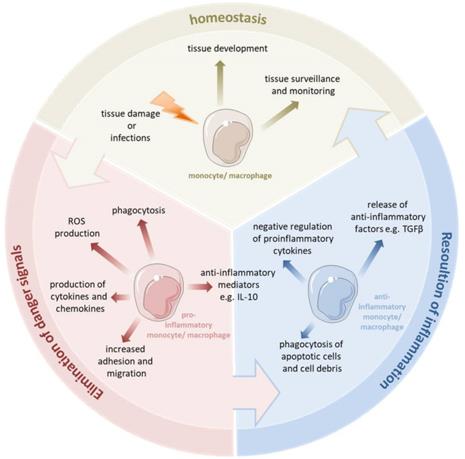
 Monocytes and macrophages are a central component of the innate immune system (Austermann J, Roth J, Barczyk-Kahlert K. 2022).
What are Macrophages?
Macrophages are specialized cells involved in the detection, phagocytosis and destruction of bacteria and other harmful organisms. In addition, they can also present antigens to T cells and initiate inflammation by releasing cytokines that activate other cells.
Macrophage Physiology
Macrophages originate from blood monocytes that leave the circulation to differentiate in different tissues. There is a substantial heterogeneity among each macrophage population, which most probably reflects the required level of specialization within the environment of any given tissue. This heterogeneity is reflected in their morphology, the type of pathogens they can recognize, as well as the levels of inflammatory cytokines they produce.
Macrophages migrate to and circulate within almost every tissue, patrolling for pathogens or clearing out dead cells. Macrophages are able to detect products of bacteria and other microorganisms using a system of recognition receptors such as Toll-like receptors (TLRs). These receptors can specifically bind to different pathogen components such as sugars (LPS), RNA, DNA or extracellular proteins. In addition, macrophages produce reactive oxygen species, such as nitric oxide, which can kill phagocytosed bacteria.
Macrophage Subpopulations
Macrophages can be roughly categorized into two main types: M1 and M2 macrophages. The M1 type, referred to as classically-activated macrophages (CAM), comprise immune effector cells with an acute inflammatory phenotype. They are highly aggressive to bacteria and produce large amounts of lymphokines. The M2 type, known as alternatively-activated macrophages (AAM), can be separated into at least three subgroups. These subtypes have diverse functions, including regulation of immunity, maintenance of tolerance, and tissue repair/ wound healing. The commonly accepted marker profile for M1 macrophages is CD68+/CD80+, while that of M2 macrophages is CD68+/CD163+.
Creative Bioarray Relevant Recommendations
Accelerate your programs with Creative Bioarray's extensive inventory and prospective network of monocytes and macrophages.
| Cat. No. | Product Name |
| CSC-7640W | Normal Human Peripheral Blood MONOCYTES(CD14+) |
| CSC-C4472X | Mobilized PB CD14+ Monocytes |
| CSC-C4490X | Frozen/Untouched Normal PB CD14+ Monocytes |
| CSC-C4613J | Human Peripheral Blood CD14 Monocytes |
| CSC-C4535X | Cord Blood CD14+ Monocytes |
| CSC-C1671 | Human Monocytes (hMo-PB) |
| CSC-C4493X | Frozen/Positively Selected Normal PB CD14+ Monocytes |
| CSC-C4715Z | M1/M2 Macrophages |
| CSC-C4427X | Normal PB Monocyte Derived Macrophages |
View our full range of macrophages and monocytes!
For research use only. Not for any other purpose.

