- You are here: Home
- Resources
- Explore & Learn
- Exosome
- Applications of MSC-EVs in Immune Regulation and Regeneration
Support
-
Cell Services
- Cell Line Authentication
- Cell Surface Marker Validation Service
-
Cell Line Testing and Assays
- Toxicology Assay
- Drug-Resistant Cell Models
- Cell Viability Assays
- Cell Proliferation Assays
- Cell Migration Assays
- Soft Agar Colony Formation Assay Service
- SRB Assay
- Cell Apoptosis Assays
- Cell Cycle Assays
- Cell Angiogenesis Assays
- DNA/RNA Extraction
- Custom Cell & Tissue Lysate Service
- Cellular Phosphorylation Assays
- Stability Testing
- Sterility Testing
- Endotoxin Detection and Removal
- Phagocytosis Assays
- Cell-Based Screening and Profiling Services
- 3D-Based Services
- Custom Cell Services
- Cell-based LNP Evaluation
-
Stem Cell Research
- iPSC Generation
- iPSC Characterization
-
iPSC Differentiation
- Neural Stem Cells Differentiation Service from iPSC
- Astrocyte Differentiation Service from iPSC
- Retinal Pigment Epithelium (RPE) Differentiation Service from iPSC
- Cardiomyocyte Differentiation Service from iPSC
- T Cell, NK Cell Differentiation Service from iPSC
- Hepatocyte Differentiation Service from iPSC
- Beta Cell Differentiation Service from iPSC
- Brain Organoid Differentiation Service from iPSC
- Cardiac Organoid Differentiation Service from iPSC
- Kidney Organoid Differentiation Service from iPSC
- GABAnergic Neuron Differentiation Service from iPSC
- Undifferentiated iPSC Detection
- iPSC Gene Editing
- iPSC Expanding Service
- MSC Services
- Stem Cell Assay Development and Screening
- Cell Immortalization
-
ISH/FISH Services
- In Situ Hybridization (ISH) & RNAscope Service
- Fluorescent In Situ Hybridization
- FISH Probe Design, Synthesis and Testing Service
-
FISH Applications
- Multicolor FISH (M-FISH) Analysis
- Chromosome Analysis of ES and iPS Cells
- RNA FISH in Plant Service
- Mouse Model and PDX Analysis (FISH)
- Cell Transplantation Analysis (FISH)
- In Situ Detection of CAR-T Cells & Oncolytic Viruses
- CAR-T/CAR-NK Target Assessment Service (ISH)
- ImmunoFISH Analysis (FISH+IHC)
- Splice Variant Analysis (FISH)
- Telomere Length Analysis (Q-FISH)
- Telomere Length Analysis (qPCR assay)
- FISH Analysis of Microorganisms
- Neoplasms FISH Analysis
- CARD-FISH for Environmental Microorganisms (FISH)
- FISH Quality Control Services
- QuantiGene Plex Assay
- Circulating Tumor Cell (CTC) FISH
- mtRNA Analysis (FISH)
- In Situ Detection of Chemokines/Cytokines
- In Situ Detection of Virus
- Transgene Mapping (FISH)
- Transgene Mapping (Locus Amplification & Sequencing)
- Stable Cell Line Genetic Stability Testing
- Genetic Stability Testing (Locus Amplification & Sequencing + ddPCR)
- Clonality Analysis Service (FISH)
- Karyotyping (G-banded) Service
- Animal Chromosome Analysis (G-banded) Service
- I-FISH Service
- AAV Biodistribution Analysis (RNA ISH)
- Molecular Karyotyping (aCGH)
- Droplet Digital PCR (ddPCR) Service
- Digital ISH Image Quantification and Statistical Analysis
- SCE (Sister Chromatid Exchange) Analysis
- Biosample Services
- Histology Services
- Exosome Research Services
- In Vitro DMPK Services
-
In Vivo DMPK Services
- Pharmacokinetic and Toxicokinetic
- PK/PD Biomarker Analysis
- Bioavailability and Bioequivalence
- Bioanalytical Package
- Metabolite Profiling and Identification
- In Vivo Toxicity Study
- Mass Balance, Excretion and Expired Air Collection
- Administration Routes and Biofluid Sampling
- Quantitative Tissue Distribution
- Target Tissue Exposure
- In Vivo Blood-Brain-Barrier Assay
- Drug Toxicity Services
Applications of MSC-EVs in Immune Regulation and Regeneration
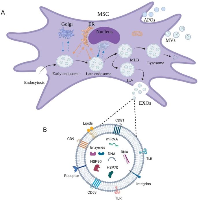
Mesenchymal stem cells (MSCs) can be widely isolated from various tissues including bone marrow, umbilical cord, and adipose tissue, with the potential for self-renewal and multipotent differentiation. Extracellular vesicles (EVs) are fundamental paracrine effectors of MSCs and play a crucial role in intercellular communication. Since MSC-derived EVs retain the function of protocells and have lower immunogenicity, they have a wide range of prospective therapeutic applications with advantages over cell therapy.
 Fig.1 The development and main types of extracellular vesicles. (Kou M, et al., 2022)
Role of EVs in Osteoarthritis
- Osteoarthritis (OA) is the principal form of joint disease with unclear pathogenesis, presenting with pain and stiffness, and in some cases, disability. Recently, MSC-EVs have been proven to have both regenerative and immunoregulatory benefits in OA, such as BMSV-EVs, hBMSC-EVs, BMSC-Exos, hASC-EVs, SMSC-EVs, SMSC-Exos, UMSC-Exos, and others.
- Several studies have reported that hBMSC-EVs play a significant role in the treatment of OA by inhibiting some pro-inflammatory pathways and factors and enhancing the proliferation and migration of chondrocytes. In addition, recent studies have also examined the effect of miRNAs in MSC-EVs.
Role of EVs in Spinal Cord Injury
Spinal cord injury (SCI) arises following damage to its structure and function by various pathogenic factors, with consequent spinal cord dysfunction including that of movement, sensation, and reflexes. Due to the limited regenerative ability of nerve components, MSC-EVs have been recently viewed as a promising clinical treatment for SCI, including BMSC-Exos, BMSV-EVs, hPMSC-Exos, MSC-EVs, and MCS-Exos.
Role of EVs in Skin Injury
Skin injury is quite common. Skin regeneration is typically accompanied by four overlapping processes, inflammation, angiogenesis, new tissue formation, and remodeling. There is recent evidence that human-derived MSC-Exos effectively benefit skin damage and accelerate wound healing by modulating related signaling pathways.
Role of EVs in Liver Fibrosis
Liver fibrosis is a pathophysiological process and refers to the abnormal proliferation of intrahepatic connective tissue due to various pathogenic factors. Recently, the use of MSC-EVs has been considered a new therapeutic approach to repair liver fibrosis. Human bone MSC-EVs inhibited expression of Wnt/β-catenin pathway components, α-SMA, and type I collagen, thereby preventing stellate cell activation and increasing hepatocyte regeneration.
Role of EVs in Kidney Fibrosis
Renal fibrosis is a gradual pathophysiological process during which kidney function progresses from healthy to injured, then to damage with an ultimate loss of function. Increasingly, MSC-EVs have been studied in the treatment of renal fibrosis using various models, including
Role of EVs in Lung Fibrosis
- Renal fibrosis is a gradual pathophysiological process during which kidney function progresses from healthy to injured, then to damage with an ultimate loss of function. Increasingly, MSC-EVs have been studied in the treatment of renal fibrosis using various models. Additionally, MSC-EVs can reverse lung injury and pulmonary fibrosis by expressing influential miRNAs.
- BMSC-Exos have been shown to significantly ameliorate hyperoxia (HYRX)-induced bronchopulmonary dysplasia (BPD), alveolar fibrosis, and pulmonary vascular remodeling by suppressing M1 macrophage production and enhancing M2 macrophage generation.
Creative Bioarray Relevant Recommendations
Creative Bioarray has developed three kinds of immortalized human mesenchymal stem cells (from various tissues, including bone marrow, placental tissue, and adipose tissue) by non-viral gene transfer of a plasmid carrying the hTERT gene, which allows the production of EVs. Apart from that, we can also provide EVs from Immortalized Human Adipose-derived Mesenchymal Stem Cells-hTERT.
Reference
- Kou M, et al. (2022). "Mesenchymal stem cell-derived extracellular vesicles for immunomodulation and regeneration: a next generation therapeutic tool?" Cell Death Dis. 13 (7), 580.
For research use only. Not for any other purpose.

