Featured Products
Our Promise to You
Guaranteed product quality, expert customer support

ONLINE INQUIRY

- Specification
- Background
- Scientific Data
- Q & A
- Customer Review
The KKU-213 cell line originated from the intrahepatic cholangiocarcinoma tissue of a 58-year-old male patient. The patient had an adenosquamous carcinoma tumor which contained granulomas as a result of Opisthorchis viverrini (liver fluke) egg infection. Cholangiocarcinoma is a malignant epithelial tumor of the bile duct which is classified as intrahepatic or extrahepatic depending on its anatomical location. The KKU-213 cell line comes from intrahepatic cholangiocarcinoma and frequently occurs with liver fluke infections. The KKU-213 cell line reacts strongly to anticancer treatments including Exemestane and Tamoxifen (TAM) which work to decrease cell growth by reducing CYP19A1 gene expression.
Researchers utilize the KKU-213 cell line to evaluate the effectiveness and action mechanisms of anticancer drugs. For instance, studies indicates that BJA-95 triggers cell death in KKU-213 cells by activating the NF-κB signaling pathway. Scientists examine specific gene functions in cholangiocarcinoma development by deleting or overexpressing miR-210, CD44, and APEX1. Furthermore, researchers use the KKU-213 cell line to understand tumor microenvironment impacts on cholangiocarcinoma cell behavior. For example, the application of IL-33 treatment results in a marked increase in both the proliferation rate and invasive abilities of KKU-213 cells.
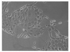 Fig. 1. Characteristics of the KKU-213 cells (Uthaisar K, Vaeteewoottacharn K, et al., 2016).
Flavopiridol Inhibited Proliferation in CCA Cell Lines and Induced G2/M Cell Cycle Arrest
Scientists recently discovered that Flavopiridol as a pan-cyclin-dependent kinase (CDK) inhibitor functions as an effective antitumor agent against multiple cancer types. Saisomboon et al. tested flavopiridol's impact on CCA cell proliferation and antitumor efficacy in a xenograft mouse model to determine its potential effectiveness for CCA treatment.
They assessed the proliferation of four CCA cell lines (KKU-055, KKU-100, KKU-213, and KKU-214) using the MTT assay after subjecting them to flavopiridol treatment at concentrations of 0, 50, 100, 200, and 300 nM across time points of 24, 48, and 72 hours. The proliferation of cells decreased significantly in response to flavopiridol treatments which showed dose- and time-dependent effects (Fig. 1A). IC50 values for flavopiridol demonstrated a range between 40 nM and 213 nM. Researchers investigated the relationship between proliferation inhibition and cell cycle arrest by applying flavopiridol to KKU-055 and KKU-213 cells followed by PI staining and flow cytometry analysis. The treatment of both cell lines with flavopiridol led to a dosage-dependent G2/M phase arrest (Fig. 1B). The levels of Cdc2 protein remained unchanged while there was a slight increase in cyclin B1 expression during the time course depicted in Fig. 1C, indicating that flavopiridol induces G2/M arrest, albeit weakly.
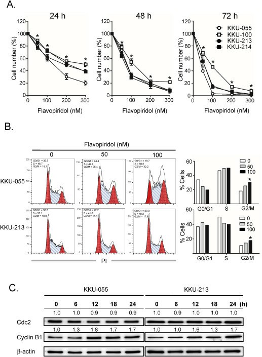 Fig. 1. (A) Flavopiridol inhibited proliferation in CCA cell lines. (B-C) Flavopiridol induced G2/M cell cycle arrest (Saisomboon S, Kariya R, et al., 2019).
Gemcitabine Sensitized CCA Cells to Enhance the Cytotoxic Effect by Stimulated T-Cells
Cholangiocarcinoma (CCA) can resist chemotherapy resulting in treatment failure. Gemcitabine, a chemotherapeutic drug, can sensitize cancer cells to become susceptible to cytotoxic T-lymphocytes (CTLs). Sawasdee et al. hypothesized that a combination of gemcitabine and CTLs would be more effective for CCA treatment than individual therapy. To test this hypothesis, they conducted an in vitro study using gemcitabine combined with CTLs to treat gemcitabine-resistant CCA (KKU-213) cells.
They examined the antitumor activity of stimulated T-cells against KKU-213 cells, measuring dead cells via Annexin V/PI staining after 24-hour co-incubation (Fig. 2A). Target cell death included PI+ (Q1), Annexin+ (Q3), and PI+/Annexin+ (Q2) populations. Results showed T-cells stimulated with SD-DC + Lys or SD-DC-PR induced over 40% cell death at a 15:1 effector-to-target (E:T) ratio, a higher effect than other conditions such as untreated DCs, unpulsed-DCs (Mo-DCs), pulsed-DCs (Mo-DCs + Lys), and SD-DCs expressing albumin (SD-DC-ALB) used as a control (Fig. 2B). To test if gemcitabine-resistant KKU-213 cells could be sensitized with a low 2 µM concentration of gemcitabine, which retains over 80% viability, cells were pretreated for 24 hours before adding stimulated T-cells at E:T ratios of 5:1, 10:1, and 15:1. After 24 hours, dead target cells were counted. Without pretreatment, T-cells stimulated by SD-DC + Lys or SD-DC-PR killed cells at rates matching the E:T ratio (Fig. 3A, B). Notably, pretreatment with gemcitabine significantly boosted T-cell killing efficiency at all E:T ratios. At 5:1, T-cells stimulated with either approach resulted in approximately 48.18 ± 8.00% (Fig. 3A) and 51.92 ± 3.84% (Fig. 3B) target cell death, both about 2.5 times higher than untreated control.
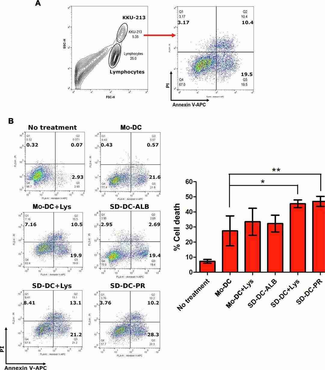 Fig. 2. Cytotoxic activities of T-cells stimulated by different types of DCs to kill KKU-213 cells (Sawasdee N, Thepmalee C, et al., 2020).
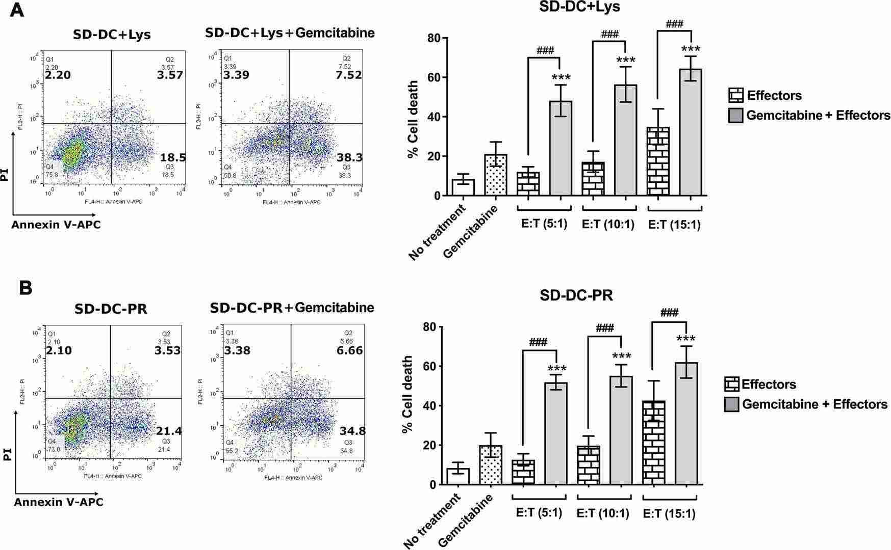 Fig. 3. Gemcitabine enhanced the cytotoxic activities of CTLs (Sawasdee N, Thepmalee C, et al., 2020).
The epithelial lining of the gall bladder consists of simple columnar cells specialized for absorption, with an apical brush border of microvilli, very similar to intestinal absorptive cells. However, unlike intestinal epithelium, gall bladder epithelium includes only this single cell type.
Ask a Question
Average Rating: 5.0 | 1 Scientist has reviewed this product
Pleased
I have been very pleased with the results I have obtained using the esophageal tumor cells from this company.
13 Nov 2022
Ease of use
After sales services
Value for money
Write your own review
- You May Also Need

Description: Japanese gallbladder carcinoma established from metastated ascite.


Description: Japanese gallbladder carcinoma from the same patient of TGBC1TKB.
Description: Japanese gallbadder carcinoma metastated to lymph node from the same patient of TGBC2TKB.

Description: Human poorly differentiated cholangiocarcinoma cell line established from biliary tract.