Featured Products
Our Promise to You
Guaranteed product quality, expert customer support

ONLINE INQUIRY
BFTC-909
Cat.No.: CSC-C2549
Species: Human
Source: kidney transitional cell carcinoma
Morphology: epithelial-like (spindle) cells growing in monolayers; cells contain considerable amounts of small, dark granules
Culture Properties: monolayer
- Specification
- Background
- Scientific Data
- Q & A
- Customer Review
Immunology: cytokeratin -, cytokeratin-7 -, cytokeratin-8 -, cytokeratin-17 -, cytokeratin-18 -
The BFTC-909 cell line derives from high-grade urothelial carcinoma of the renal pelvis in a Taiwanese patient and is recognized for its high malignant and invasive properties. This cell line forms an epithelial-like (spindle-shaped) monolayer with many small dark granules inside its cells that indicate its renal epithelial tissue origin along with the typical features of renal tubular transitional cell carcinoma. Research shows that chromosomes 5, 7, and 12 have hypermethylated DNA regions within the PCDH, HOXA, and HOXC gene clusters. The enhanced expression of the INHBA gene in BFTC-909 cells correlates with their proliferation and migration capabilities because silencing this gene through siRNA results in significant inhibition of these functions.
The BFTC-909 cell line serves as a vital research tool in urothelial carcinoma studies which encompasses drug screening and anti-tumor evaluations (such as testing the combined effects of AZD-1775 and cisplatin) gene function research (investigating cancer progression determinants like INHBA and FGFR3) and molecular mechanism investigations (exploring urothelial carcinoma mechanisms through DNA methylation patterns and gene expression profiling).
AF Interferes with Cell Cycle Progression in HT 1376 and BFTC 909 Cells
Urothelial carcinoma (UC) remains a prevalent urinary tract cancer with low five-year survival rates because patients develop resistance to cisplatin and experience cancer recurrence following surgical intervention. Drugs that generate reactive oxygen species (ROS) show potential because cancer cells with high ROS concentrations become susceptible to oxidative stress. Auranofin (AF), which works as an antirheumatic drug, shows anticancer potential through redox balance disruption. The effects of AF treatment on cell cycle progression were assessed by applying 1.5 and 3 µM AF to HT 1376 cells and 2 and 4 µM AF to BFTC 909 cells followed by flow cytometry analysis. Treatment of HT 1376 cells with AF resulted in a G0/G1 phase arrest which was evidenced by the concentration-dependent growth in the number of cells within the G0/G1 phase (Fig. 1A). The G0/G1 phase cell percentage rose from 66.06% to 71.05% at 8 hours while treatment with AF concentrations of 1.5 and 3 µM and then increased from 65.47% to 69.74% at 12 hours and from 57.00% to 65.67% at 24 hours (Fig. 2B). The application of AF resulted in increased levels of p21 and p27 while simultaneously decreasing CDK2, CDK4, cyclin D1, and cyclin E2 in HT 1376 cells (Fig. 2C) AF treatment leads to G0/G1 arrest in the examined cells.
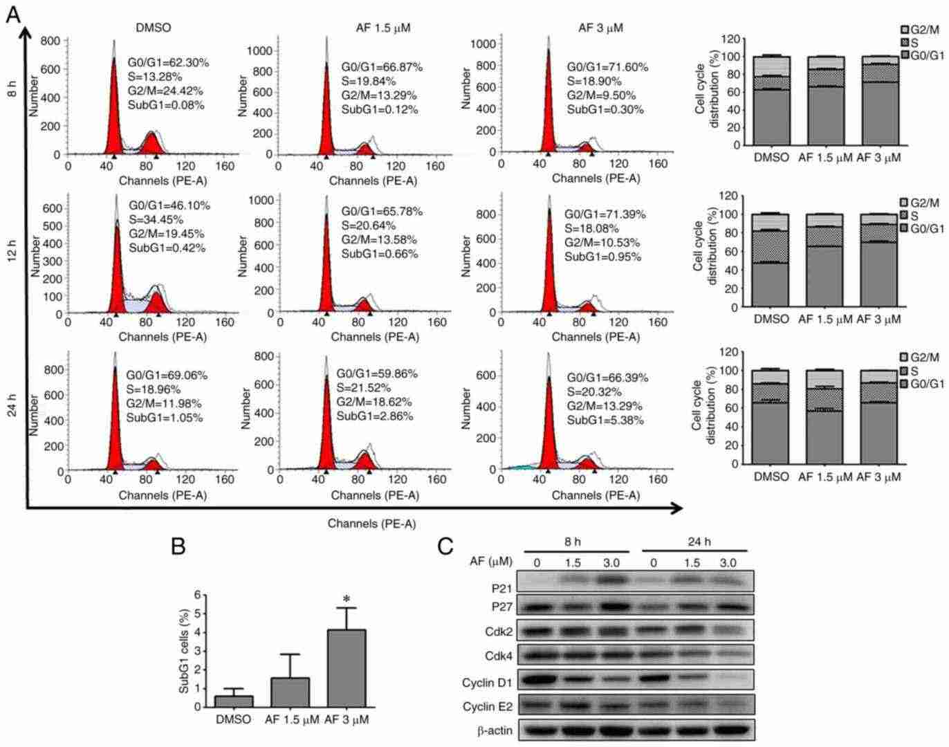 Fig. 1. AF induces G0/G1 cell cycle arrest in HT 1376 urothelial carcinoma cells (Chen S, Chao C, et al., 2021).
Downregulation of GAL1 in UC Cell Lines
UTUC, though rare, is more aggressive than bladder cancer and particularly prevalent in Taiwan. Galectin-1 (GAL1), a protein involved in tumorigenesis, is linked to poorer outcomes in various cancers, but its full role in UTUC is unexplored. This study aims to uncover GAL1's impact on UTUC survival and its oncological pathways. In vitro, GAL1's effect on tumor growth and metastasis was evaluated across four UC cell lines (BFTC-909, T24, J82, and RT4). Figure 2A, B shows that GAL1 mRNA levels were significantly high in BFTC-909, T24, and J82 cells, correlating with elevated GAL1 protein levels, while RT4 cells showed low expression. We chose BFTC-909 and T24, with the highest GAL1 levels, to create GAL1 knockdown lines (sh-Gal1). Figure 2C, D demonstrates significantly reduced GAL1 protein and mRNA in sh-GAL1-T24 and sh-GAL1-BFTC-909 cells, confirming effective LGALS1 gene silencing. Stable knockdown lines were established via lentivirus transfection. Figure 3E–H presents that GAL1 protein and mRNA were notably downregulated in sh-GAL1-RNAi-A and sh-GAL1-RNAi-B BFTC-909 and T24 cells, with RNAi-A achieving higher knockdown efficiency than RNAi-B.
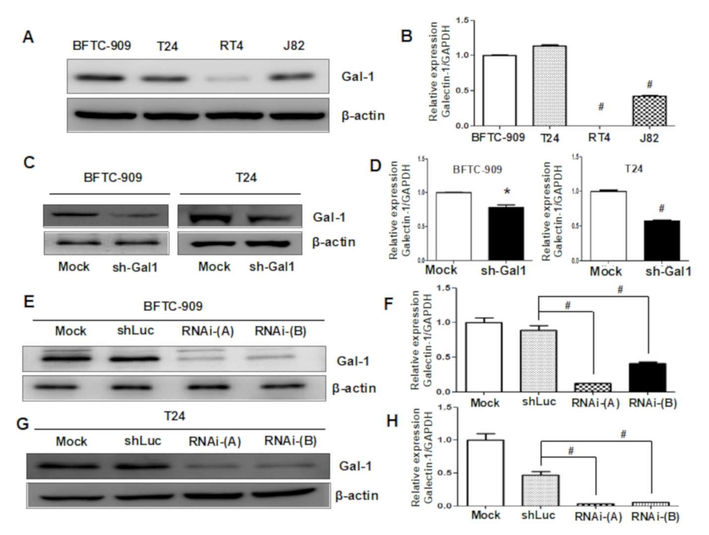 Fig. 2. Evaluation of GAL1 expression in UC cell lines and verification of downregulation efficiency through Western blotting and q-PCR analyses (Su Y-L, Luo H-L, et al., 2020).
Kidney cancer begins when healthy cells in 1 or both kidneys change and grow out of control, forming a mass called a renal cortical tumor. A tumor can be malignant, indolent, or benign. A malignant tumor is cancerous, meaning it can grow and spread to other parts of the body.
Ask a Question
Average Rating: 5.0 | 1 Scientist has reviewed this product
Excellent
I have found Creative Bioarray's cell products to be an excellent resource in my research.
03 Sep 2022
Ease of use
After sales services
Value for money
Write your own review
- You May Also Need

Description: Established from the malignant pleural effusion of a 75-year-old man with kidney carcinoma in 1987

Description: Species: human - female, 9 months old, Caucasian
Isoenzyme: G6PD, B
Karyology: diploid
Histopathology: leiomyoblastoma, renal
Note: the line will grow in soft agar

Description: Renal tumor cell from a Japanese. Transplantable to nude mice.

Description: OACP4 C was established from a primary tumour located in gastric cardia of a 55 year-old Caucasian male. The patient showed lymph node involvement with pleural metastases. OACP4 C was found to be tumourigenic in NMRI (female nude mice or NOD-SCID mice. The Y chromosome could not be detected in this cell line by short tandem repeat (STR)-PCR analysi. It is a known phenomenon that due to the increased genetic instability of cancer cell lines the Y chromosome can be rearranged or lost resulting in lack of detection.