What Are Myeloid Cell Markers?
Myeloid cells are a diverse group of immune cells originating from the myeloid lineage of hematopoietic stem cells. They play pivotal roles in various physiological and pathological processes, including immune response, inflammation, and cancer. Identifying and characterizing myeloid cell markers are essential for understanding their functions and interactions within the immune system.
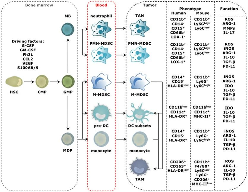
 Fig.1 The distinct steps in the progression from hematopoietic stem cells (HSCs) to tumor-infiltrating myeloid cells (TIMs) occur at different locations and start with amplification and differentiation of the HSC and its progenitors, including the common myeloid progenitor (CMP), granulocyte-monocyte progenitor (GMP), myeloblast (MB), and monocyte-dendritic cell progenitor (MDP) in the bone marrow. (Awad RM, et al., 2018)
Different Types of Myeloid Cells
Myeloid cells encompass various cell types, each with unique functions and characteristics. This lineage includes various cell types, each with unique functions and characteristics. Among the most prominent myeloid cell types are macrophages, neutrophils, dendritic cells (DCs), and myeloid-derived suppressor cells (MDSCs).
- Macrophages are versatile phagocytic cells that play a crucial role in innate immunity, pathogen clearance, and tissue homeostasis.
- Neutrophils are the most abundant type of white blood cells and are primarily responsible for the initial immune response against infectious agents.
- DCs are professional antigen-presenting cells that bridge the gap between innate and adaptive immunity, playing a pivotal role in initiating and shaping the adaptive immune response.
- MDSCs are a heterogeneous population of immature myeloid cells that have the unique ability to suppress T cell responses, making them an important player in the tumor microenvironment.
How to Identify and Characterize Myeloid Cell Markers?
Myeloid cell identification and characterization rely heavily on the expression of specific cell surface markers, also known as cluster of differentiation (CD) antigens. These markers serve as unique identifiers for the various myeloid cell subtypes, allowing researchers and clinicians to distinguish and study them in depth. The identification and characterization of myeloid cell markers involve a combination of techniques, including flow cytometry, immunohistochemistry (IHC), and gene expression profiling.
- Flow cytometry. It is a powerful tool that enables the simultaneous analysis of multiple cell surface markers on individual cells. Researchers can accurately identify and quantify the different myeloid cell populations within a heterogeneous sample by staining cells with fluorescently labeled antibodies targeting specific myeloid cell markers.
- IHC. This method allows researchers to study the spatial distribution and tissue-specific expression patterns of myeloid cell markers, providing valuable insights into the role of these cells in the context of their native microenvironment. Using a range of myeloid cell-specific antibodies and advanced microscopy techniques, scientists can precisely map the presence and localization of markers like CD11b, CD14, and CD163 within tumor tissues, lymphoid organs, or other relevant biological samples.
- Gene expression profiling. It provides insights into the molecular characteristics of myeloid cells that help identify markers that are unique or differentially expressed in specific cell types or states. By analyzing the expression patterns of key myeloid cell marker genes, researchers can gain insights into the developmental origins, activation states, and potential functional roles of different myeloid cell populations.
Common Myeloid Cell Markers
- CD11b, also known as integrin αM, is a hallmark marker of myeloid cells. It is expressed on the surface of monocytes, macrophages, granulocytes, and MDSCs. As a member of the integrin family, CD11b plays a crucial role in cell-cell and cell-extracellular matrix interactions, facilitating the migration and adhesion of myeloid cells to sites of inflammation or infection.
- CD14 is a glycosylphosphatidylinositol (GPI)-anchored protein that serves as a co-receptor for the detection of bacterial lipopolysaccharide (LPS). It is primarily expressed on the surface of monocytes and macrophages, and its upregulation is associated with the differentiation and activation of these myeloid cells. CD14 is a valuable marker for identifying and characterizing the monocyte/macrophage lineage.
- CD16, known as FcγRIII, is a low-affinity Fc receptor for IgG, expressed on the surface of neutrophils, natural killer cells, and a subset of monocytes.
- CD33, also known as Siglec-3, is a member of the sialic acid-binding immunoglobulin-like lectin (Siglec) family. It is expressed on the surface of myeloid progenitor cells, monocytes, and a subset of DCs. CD33 plays a role in the regulation of myeloid cell function and has been the target of various therapeutic approaches, particularly in the treatment of myeloid-derived malignancies.
- CD64, also known as FcγRI, is a high-affinity receptor for the Fc portion of immunoglobulin G (IgG). It is primarily expressed on the surface of mature monocytes, macrophages, and DCs, and its expression is upregulated in response to inflammatory stimuli. CD64 is a valuable marker for identifying and quantifying activated myeloid cells, particularly in the context of infectious diseases and autoimmune disorders.
- CD115, known as CSF-1R, is the receptor for macrophage colony-stimulating factor (M-CSF), expressed on the surface of monocytes, macrophages, and their progenitors.
Myeloid Cell Markers in Cancer Research
Myeloid cells, particularly their subtypes, have garnered significant attention in the field of cancer research due to their pivotal roles in the tumor microenvironment. Understanding the expression and function of myeloid cell markers has become crucial for elucidating the complex interactions between the immune system and cancer.
MDSC markers in cancer
MDSCs are a heterogeneous population of immature myeloid cells that can suppress T-cell responses, thereby promoting tumor growth and metastasis. Key MDSC markers include CD11b, CD33, CD14, and CD15, which are often used in combination to identify and characterize these cells in the context of cancer.
Macrophage markers in cancer
Tumor-associated macrophages (TAMs) are a crucial component of the tumor microenvironment and can exhibit both pro-tumorigenic and anti-tumorigenic properties, depending on their polarization state. Commonly used macrophage markers in cancer research include CD11b, CD68, CD163, and CD206, which help distinguish between different macrophage subsets and their functional roles.
Neutrophil markers in cancer
Neutrophils, while traditionally associated with the initial immune response against pathogens, have also been implicated in various aspects of cancer progression, including tumor angiogenesis and metastasis. Neutrophil markers such as CD11b, CD66b, and CD16 are frequently used to identify and study the role of these cells in the tumor microenvironment.
Dendritic cells in cancer
DCs play a critical role in antigen presentation and the initiation of adaptive immune responses against cancer cells. Markers like CD11c, CD1a, and HLA-DR are commonly used to identify and characterize dendritic cell populations in the context of cancer immunotherapy and tumor immune surveillance.
Creative Bioarray Relevant Recommendations
| Cat. No. | Product Name |
| CSC-C4573X | Bone Marrow CD33+ Myeloid Cells |
| CSC-C4618J | Human Bone Marrow CD33+CD66+ Myeloid Cells |
| CSK-CI065 | SuperBeads® Human Myeloid Cells Isolation Kit |
Reference
- Awad RM, et al. (2018). "Turn Back the TIMe: Targeting Tumor Infiltrating Myeloid Cells to Revert Cancer Progression." Front Immunol. 9: 1977.

