- You are here: Home
- Resources
- Explore & Learn
- Cell Biology
- Multi-Differentiation of Peripheral Blood Mononuclear Cells
Support
-
Cell Services
- Cell Line Authentication
- Cell Surface Marker Validation Service
-
Cell Line Testing and Assays
- Toxicology Assay
- Drug-Resistant Cell Models
- Cell Viability Assays
- Cell Proliferation Assays
- Cell Migration Assays
- Soft Agar Colony Formation Assay Service
- SRB Assay
- Cell Apoptosis Assays
- Cell Cycle Assays
- Cell Angiogenesis Assays
- DNA/RNA Extraction
- Custom Cell & Tissue Lysate Service
- Cellular Phosphorylation Assays
- Stability Testing
- Sterility Testing
- Endotoxin Detection and Removal
- Phagocytosis Assays
- Cell-Based Screening and Profiling Services
- 3D-Based Services
- Custom Cell Services
- Cell-based LNP Evaluation
-
Stem Cell Research
- iPSC Generation
- iPSC Characterization
-
iPSC Differentiation
- Neural Stem Cells Differentiation Service from iPSC
- Astrocyte Differentiation Service from iPSC
- Retinal Pigment Epithelium (RPE) Differentiation Service from iPSC
- Cardiomyocyte Differentiation Service from iPSC
- T Cell, NK Cell Differentiation Service from iPSC
- Hepatocyte Differentiation Service from iPSC
- Beta Cell Differentiation Service from iPSC
- Brain Organoid Differentiation Service from iPSC
- Cardiac Organoid Differentiation Service from iPSC
- Kidney Organoid Differentiation Service from iPSC
- GABAnergic Neuron Differentiation Service from iPSC
- Undifferentiated iPSC Detection
- iPSC Gene Editing
- iPSC Expanding Service
- MSC Services
- Stem Cell Assay Development and Screening
- Cell Immortalization
-
ISH/FISH Services
- In Situ Hybridization (ISH) & RNAscope Service
- Fluorescent In Situ Hybridization
- FISH Probe Design, Synthesis and Testing Service
-
FISH Applications
- Multicolor FISH (M-FISH) Analysis
- Chromosome Analysis of ES and iPS Cells
- RNA FISH in Plant Service
- Mouse Model and PDX Analysis (FISH)
- Cell Transplantation Analysis (FISH)
- In Situ Detection of CAR-T Cells & Oncolytic Viruses
- CAR-T/CAR-NK Target Assessment Service (ISH)
- ImmunoFISH Analysis (FISH+IHC)
- Splice Variant Analysis (FISH)
- Telomere Length Analysis (Q-FISH)
- Telomere Length Analysis (qPCR assay)
- FISH Analysis of Microorganisms
- Neoplasms FISH Analysis
- CARD-FISH for Environmental Microorganisms (FISH)
- FISH Quality Control Services
- QuantiGene Plex Assay
- Circulating Tumor Cell (CTC) FISH
- mtRNA Analysis (FISH)
- In Situ Detection of Chemokines/Cytokines
- In Situ Detection of Virus
- Transgene Mapping (FISH)
- Transgene Mapping (Locus Amplification & Sequencing)
- Stable Cell Line Genetic Stability Testing
- Genetic Stability Testing (Locus Amplification & Sequencing + ddPCR)
- Clonality Analysis Service (FISH)
- Karyotyping (G-banded) Service
- Animal Chromosome Analysis (G-banded) Service
- I-FISH Service
- AAV Biodistribution Analysis (RNA ISH)
- Molecular Karyotyping (aCGH)
- Droplet Digital PCR (ddPCR) Service
- Digital ISH Image Quantification and Statistical Analysis
- SCE (Sister Chromatid Exchange) Analysis
- Biosample Services
- Histology Services
- Exosome Research Services
- In Vitro DMPK Services
-
In Vivo DMPK Services
- Pharmacokinetic and Toxicokinetic
- PK/PD Biomarker Analysis
- Bioavailability and Bioequivalence
- Bioanalytical Package
- Metabolite Profiling and Identification
- In Vivo Toxicity Study
- Mass Balance, Excretion and Expired Air Collection
- Administration Routes and Biofluid Sampling
- Quantitative Tissue Distribution
- Target Tissue Exposure
- In Vivo Blood-Brain-Barrier Assay
- Drug Toxicity Services
Multi-Differentiation of Peripheral Blood Mononuclear Cells
Peripheral blood mononuclear cells (PBMCs) are a diverse group of immune cells that play a crucial role in the body's immune response. They include lymphocytes (B cells, T cells, and natural killer cells), monocytes, and dendritic cells. PBMCs have been reported to contain a multitude of distinct multipotent progenitor cell populations and possess the potential to differentiate into blood cells, endothelial cells, hepatocytes, cardiomyogenic cells, smooth muscle cells, osteoblasts, osteoclasts, epithelial cells, neural cells, or myofibroblasts under appropriate conditions.
Multi-Differentiation of PBMCs
- Differentiation of PBMCs into blood cells
PBMCs can differentiate into various types of blood cells, including red blood cells (erythrocytes), white blood cells (leukocytes), and platelets. Human PBMCs can also produce other hematopoietic colonies, including colony-forming unit-granulocyte-erythroid monocyte-macrophage-megakaryocyte, colony-forming unit monocyte-macrophage, blast-forming unit-erythroid and colony-forming unit-erythroid precursors. - Differentiation of PBMCs into endothelial cells
Endothelial cells form the inner lining of blood vessels and play a critical role in angiogenesis and vascular repair. PBMCs can be differentiated into endothelial cells through a combination of growth factors and culture conditions. This process mimics the natural development of endothelial cells from precursor cells during embryogenesis. - Differentiation of PBMCs into hepatocytes
Human PBMCs have the potential to differentiate into hepatocyte-like cells in vitro in the presence of hepatocyte growth factor or fibroblast growth factor (FGF)-4. During induction culture, cells gradually rounded but did not assume the polygonal shape of mature hepatocytes, which might suggest additional signals are required for full differentiation. - Differentiation of PBMCs into muscle cells
Growth factors such as transforming growth factor and platelet-derived growth factor-BB participate in smooth muscle cell (SMC) differentiation. Human PBMCs grown in platelet-derived growth factor BB-enriched medium differentiated into smooth muscle outgrowth cells that were positive for SMC-specific actin (SMA), myosin heavy chain, and calponin at both the mRNA and protein levels. - Differentiation of PBMCs into neural cells
The nervous system is composed of neurons and glial cells, which coordinate and regulate various physiological processes. PBMCs can differentiate into neural cells, including neurons and astrocytes, through the activation of specific signaling pathways and culture conditions.
Methods for the Isolation and Purification of PBMCs
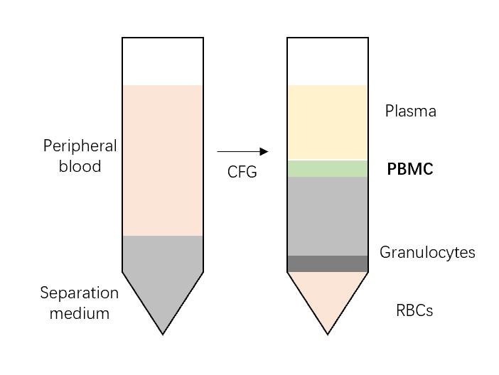
 Fig. 1 Schematic diagram for separation of PBMC.
- The isolation and purification of PBMCs from peripheral blood are crucial steps for downstream applications. Several methods are commonly used for PBMC isolation, including density gradient centrifugation, magnetic bead separation, and microfluidic techniques.
- In density gradient centrifugation, blood is layered on top of a density gradient medium, and centrifugation separates the PBMCs from other blood components based on their density. Magnetic bead separation utilizes specific antibody-coated magnetic beads to selectively bind and isolate PBMCs from other cell types. Microfluidic techniques offer a more automated and high-throughput approach for PBMC isolation.
Creative Bioarray Relevant Recommendations
Creative Bioarray provides a high-quality and comprehensive range of PBMCs, to accelerate our customers' scientific research. Specifically, our products include but are not limited to Human Normal Peripheral Blood Mononuclear Cells (PBMCs), Human MULTIPLE MYELOMA, Peripheral Blood Mononuclear Cells, and Human Peripheral Blood Mononuclear Cells (HPBMC).
For research use only. Not for any other purpose.

