- You are here: Home
- Resources
- Explore & Learn
- Histology
- Mitochondrial Staining
Support
-
Cell Services
- Cell Line Authentication
- Cell Surface Marker Validation Service
-
Cell Line Testing and Assays
- Toxicology Assay
- Drug-Resistant Cell Models
- Cell Viability Assays
- Cell Proliferation Assays
- Cell Migration Assays
- Soft Agar Colony Formation Assay Service
- SRB Assay
- Cell Apoptosis Assays
- Cell Cycle Assays
- Cell Angiogenesis Assays
- DNA/RNA Extraction
- Custom Cell & Tissue Lysate Service
- Cellular Phosphorylation Assays
- Stability Testing
- Sterility Testing
- Endotoxin Detection and Removal
- Phagocytosis Assays
- Cell-Based Screening and Profiling Services
- 3D-Based Services
- Custom Cell Services
- Cell-based LNP Evaluation
-
Stem Cell Research
- iPSC Generation
- iPSC Characterization
-
iPSC Differentiation
- Neural Stem Cells Differentiation Service from iPSC
- Astrocyte Differentiation Service from iPSC
- Retinal Pigment Epithelium (RPE) Differentiation Service from iPSC
- Cardiomyocyte Differentiation Service from iPSC
- T Cell, NK Cell Differentiation Service from iPSC
- Hepatocyte Differentiation Service from iPSC
- Beta Cell Differentiation Service from iPSC
- Brain Organoid Differentiation Service from iPSC
- Cardiac Organoid Differentiation Service from iPSC
- Kidney Organoid Differentiation Service from iPSC
- GABAnergic Neuron Differentiation Service from iPSC
- Undifferentiated iPSC Detection
- iPSC Gene Editing
- iPSC Expanding Service
- MSC Services
- Stem Cell Assay Development and Screening
- Cell Immortalization
-
ISH/FISH Services
- In Situ Hybridization (ISH) & RNAscope Service
- Fluorescent In Situ Hybridization
- FISH Probe Design, Synthesis and Testing Service
-
FISH Applications
- Multicolor FISH (M-FISH) Analysis
- Chromosome Analysis of ES and iPS Cells
- RNA FISH in Plant Service
- Mouse Model and PDX Analysis (FISH)
- Cell Transplantation Analysis (FISH)
- In Situ Detection of CAR-T Cells & Oncolytic Viruses
- CAR-T/CAR-NK Target Assessment Service (ISH)
- ImmunoFISH Analysis (FISH+IHC)
- Splice Variant Analysis (FISH)
- Telomere Length Analysis (Q-FISH)
- Telomere Length Analysis (qPCR assay)
- FISH Analysis of Microorganisms
- Neoplasms FISH Analysis
- CARD-FISH for Environmental Microorganisms (FISH)
- FISH Quality Control Services
- QuantiGene Plex Assay
- Circulating Tumor Cell (CTC) FISH
- mtRNA Analysis (FISH)
- In Situ Detection of Chemokines/Cytokines
- In Situ Detection of Virus
- Transgene Mapping (FISH)
- Transgene Mapping (Locus Amplification & Sequencing)
- Stable Cell Line Genetic Stability Testing
- Genetic Stability Testing (Locus Amplification & Sequencing + ddPCR)
- Clonality Analysis Service (FISH)
- Karyotyping (G-banded) Service
- Animal Chromosome Analysis (G-banded) Service
- I-FISH Service
- AAV Biodistribution Analysis (RNA ISH)
- Molecular Karyotyping (aCGH)
- Droplet Digital PCR (ddPCR) Service
- Digital ISH Image Quantification and Statistical Analysis
- SCE (Sister Chromatid Exchange) Analysis
- Biosample Services
- Histology Services
- Exosome Research Services
- In Vitro DMPK Services
-
In Vivo DMPK Services
- Pharmacokinetic and Toxicokinetic
- PK/PD Biomarker Analysis
- Bioavailability and Bioequivalence
- Bioanalytical Package
- Metabolite Profiling and Identification
- In Vivo Toxicity Study
- Mass Balance, Excretion and Expired Air Collection
- Administration Routes and Biofluid Sampling
- Quantitative Tissue Distribution
- Target Tissue Exposure
- In Vivo Blood-Brain-Barrier Assay
- Drug Toxicity Services
Mitochondrial Staining
The study of mitochondria is a topic of great interest in biology due to their pivotal role in cellular function. Mitochondria serve as the powerhouse of the cell and are involved in various crucial cellular processes, including energy production and apoptosis. To effectively study and visualize the structure and functions of mitochondria, researchers often use a technique known as mitochondrial staining.

What Is Mitochondrial Staining?
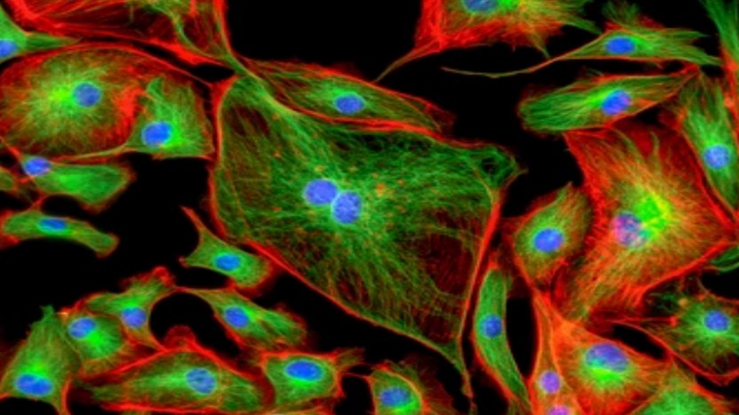
Mitochondrial staining is a technique used to selectively target and visualize mitochondria within a cell or tissue sample. By utilizing specific dyes or stains, researchers can gain insights into the morphology, distribution, and activity of mitochondria in various biological contexts.
Types of Mitochondrial Stains
- Fluorescent Dyes. Fluorescent dyes such as MitoTracker dyes are commonly used for mitochondrial staining. These dyes selectively accumulate in active mitochondria, emitting a bright fluorescence that can be visualized using fluorescence microscopy. They are available in various colors, allowing for multiplexing and co-localization studies.
- Rhodamine 123. It is another widely used fluorescent stain for mitochondria. It accumulates in mitochondria based on their membrane potential, providing insights into mitochondrial function and health.
- JC-1. It is a cationic dye that accumulates in mitochondria in a membrane potential-dependent manner. It exhibits potential-dependent accumulation in mitochondria, allowing for the assessment of mitochondrial membrane potential.
- MitoSOX Red. It is a selective fluorogenic dye for mitochondria, often used to assess mitochondrial superoxide levels.
Mechanism of Mitochondrial Staining
The mechanism of mitochondrial staining varies based on the specific stain used. However, in general, these stains are designed to selectively target mitochondria based on certain properties such as membrane potential, activity, or mitochondrial-specific components, allowing for their visualization under a fluorescent microscope or other imaging techniques.
Applications of Mitochondrial Staining
- Cell viability and health assessment. Mitochondrial staining is commonly used to assess cell viability and health. Changes in mitochondrial morphology, membrane potential, or superoxide levels can provide valuable insights into the cell's physiological state.
- Mitochondrial dynamics. By visualizing mitochondria using specific stains, researchers can study mitochondrial dynamics including fusion, fission, and movement within the cell. This is crucial for understanding the regulation of mitochondrial morphology and distribution.
- Pharmacological studies. Mitochondrial staining is used by researchers to evaluate the impact of drugs and compounds on mitochondrial function. This is crucial for assessing potential therapeutics and comprehending their effects on cellular metabolism and energy production.
Creative Bioarray Relevant Recommendations
Creative Bioarray provides a diverse range of advanced mitochondrial dyes. Our comprehensive selection of dyes, including fluorescent probes and stains, enables precise and reliable visualization of mitochondria, contributing to the advancement of mitochondrial research across various fields of biology and medicine.
| Cat. No. | Product Name |
| FDIR-D0102 | VitalTracker® Blue Mitochondria Dye |
| FDIR-D0104 | JC-1 |
| FDIR-D0105 | JC-10 |
| FDIR-D0106 | Rhodamine B |
| FDIR-D0107 | VitalTracker® Red Mitochondria Dye |
For research use only. Not for any other purpose.

