- You are here: Home
- Resources
- Explore & Learn
- Cell Biology
- Human Primary Cells: Definition, Assay, Applications
Support
-
Cell Services
- Cell Line Authentication
- Cell Surface Marker Validation Service
-
Cell Line Testing and Assays
- Toxicology Assay
- Drug-Resistant Cell Models
- Cell Viability Assays
- Cell Proliferation Assays
- Cell Migration Assays
- Soft Agar Colony Formation Assay Service
- SRB Assay
- Cell Apoptosis Assays
- Cell Cycle Assays
- Cell Angiogenesis Assays
- DNA/RNA Extraction
- Custom Cell & Tissue Lysate Service
- Cellular Phosphorylation Assays
- Stability Testing
- Sterility Testing
- Endotoxin Detection and Removal
- Phagocytosis Assays
- Cell-Based Screening and Profiling Services
- 3D-Based Services
- Custom Cell Services
- Cell-based LNP Evaluation
-
Stem Cell Research
- iPSC Generation
- iPSC Characterization
-
iPSC Differentiation
- Neural Stem Cells Differentiation Service from iPSC
- Astrocyte Differentiation Service from iPSC
- Retinal Pigment Epithelium (RPE) Differentiation Service from iPSC
- Cardiomyocyte Differentiation Service from iPSC
- T Cell, NK Cell Differentiation Service from iPSC
- Hepatocyte Differentiation Service from iPSC
- Beta Cell Differentiation Service from iPSC
- Brain Organoid Differentiation Service from iPSC
- Cardiac Organoid Differentiation Service from iPSC
- Kidney Organoid Differentiation Service from iPSC
- GABAnergic Neuron Differentiation Service from iPSC
- Undifferentiated iPSC Detection
- iPSC Gene Editing
- iPSC Expanding Service
- MSC Services
- Stem Cell Assay Development and Screening
- Cell Immortalization
-
ISH/FISH Services
- In Situ Hybridization (ISH) & RNAscope Service
- Fluorescent In Situ Hybridization
- FISH Probe Design, Synthesis and Testing Service
-
FISH Applications
- Multicolor FISH (M-FISH) Analysis
- Chromosome Analysis of ES and iPS Cells
- RNA FISH in Plant Service
- Mouse Model and PDX Analysis (FISH)
- Cell Transplantation Analysis (FISH)
- In Situ Detection of CAR-T Cells & Oncolytic Viruses
- CAR-T/CAR-NK Target Assessment Service (ISH)
- ImmunoFISH Analysis (FISH+IHC)
- Splice Variant Analysis (FISH)
- Telomere Length Analysis (Q-FISH)
- Telomere Length Analysis (qPCR assay)
- FISH Analysis of Microorganisms
- Neoplasms FISH Analysis
- CARD-FISH for Environmental Microorganisms (FISH)
- FISH Quality Control Services
- QuantiGene Plex Assay
- Circulating Tumor Cell (CTC) FISH
- mtRNA Analysis (FISH)
- In Situ Detection of Chemokines/Cytokines
- In Situ Detection of Virus
- Transgene Mapping (FISH)
- Transgene Mapping (Locus Amplification & Sequencing)
- Stable Cell Line Genetic Stability Testing
- Genetic Stability Testing (Locus Amplification & Sequencing + ddPCR)
- Clonality Analysis Service (FISH)
- Karyotyping (G-banded) Service
- Animal Chromosome Analysis (G-banded) Service
- I-FISH Service
- AAV Biodistribution Analysis (RNA ISH)
- Molecular Karyotyping (aCGH)
- Droplet Digital PCR (ddPCR) Service
- Digital ISH Image Quantification and Statistical Analysis
- SCE (Sister Chromatid Exchange) Analysis
- Biosample Services
- Histology Services
- Exosome Research Services
- In Vitro DMPK Services
-
In Vivo DMPK Services
- Pharmacokinetic and Toxicokinetic
- PK/PD Biomarker Analysis
- Bioavailability and Bioequivalence
- Bioanalytical Package
- Metabolite Profiling and Identification
- In Vivo Toxicity Study
- Mass Balance, Excretion and Expired Air Collection
- Administration Routes and Biofluid Sampling
- Quantitative Tissue Distribution
- Target Tissue Exposure
- In Vivo Blood-Brain-Barrier Assay
- Drug Toxicity Services
Human Primary Cells: Definition, Assay, Applications

Human primary cells play a crucial role in advancing our understanding of human biology and disease. They are isolated directly from human tissues and retain their original characteristics, providing a more accurate representation of in vivo physiology compared to immortalized cell lines.
What Are Primary Cells?
Human primary cells refer to cells that are isolated directly from human tissues and cultured in vitro for study. They are different from cell lines, which are derived from cancerous or immortalized cells that can grow indefinitely. Primary cells closely resemble their tissue of origin in terms of morphology, physiological functions, and gene expression patterns. They provide a highly valuable resource for studying human biology, disease mechanisms, and drug discovery.
Assay Methods
- Immunofluorescence staining
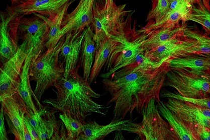
Immunofluorescence staining is commonly used to detect specific proteins within human primary cells. By utilizing antibodies labeled with fluorescent dyes, researchers can visualize the cellular localization and expression levels of target proteins. This technique helps in identifying cell subpopulations, studying protein-protein interactions, and investigating changes in protein expression during disease progression. - Flow cytometry
Flow cytometry enables the rapid analysis of individual cells in a heterogeneous population. It utilizes fluorescently labeled antibodies or dyes to quantify and analyze cellular characteristics, such as surface markers, DNA content, and intracellular signaling molecules. Flow cytometry is particularly useful in studying immune cell populations, identifying rare cell populations, and evaluating cell cycle progression.
Applications
- Drug discovery and development
Human primary cells serve as a reliable model for evaluating the efficacy and safety of potential drugs. They can be used to screen drug candidates, study drug metabolism and toxicity, and identify personalized therapeutic approaches. - Disease research
Studying human primary cells provides valuable insights into the mechanisms underlying various diseases. Researchers can investigate cellular interactions, molecular pathways, and genetic alterations associated with diseases such as cancer, cardiovascular disorders, neurodegenerative diseases, and infectious diseases. - Tissue engineering and regenerative medicine
Human primary cells are crucial in the field of tissue engineering and regenerative medicine. By combining cells with suitable scaffolds and growth factors, researchers aim to develop functional tissues and organs for transplantation. Primary cells derived from specific tissue types, such as skin or bone, can be used to regenerate damaged tissues and promote tissue repair.
Creative Bioarray Relevant Recommendations
With a strong partner network, Creative Bioarray offers more than 1000 cell types together with selective cell culture media, suitable for various tissues. This diverse range of cell types enables scientists to address a wide array of research questions.
| Product Types | Description |
| Bone Marrow Cells | Bone marrow cells are the cells contained within the bone marrow. These include stromal cells, which are not directly involved in hematopoiesis, and hematopoietic stem cells, which are responsible for the production of blood cells such as leukocytes, erythrocytes, and platelets. |
| Peripheral Blood Mononuclear Cells | Peripheral blood mononuclear cells (PBMCs) are cells with a single, round nucleus and are collected from the peripheral or circulating blood by density centrifugation with Ficoll, a polysaccharide. |
| Umbilical Cord Cells | The human umbilical cord is a conduit between the developing embryo and the placenta. We can offer many types of umbilical cord cells for researchers to find out the relationship between abnormal cell proliferation within the umbilical cord system and the development of umbilical cord abnormalities. |
| Other Human Primary Cells | Creative Bioarray is a trusted provider of human primary cells for researchers worldwide. We have multiple cell types currently in development, so if you do not see a cell type that meets your needs, please let us know and we will work to accommodate your request. |
For research use only. Not for any other purpose.

