How to Isolate and Analyze Tumor-Infiltrating Leukocytes?
Tumor-infiltrating leukocytes (TILs) are critical components of the immune response within the tumor microenvironment. Their presence and activity can significantly influence tumor progression and patient outcomes. The ability to isolate and analyze TILs is essential for advancing our understanding of cancer immunology and developing novel therapeutic strategies.
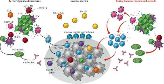
 Fig.1 TILs in the immunotherapy. (Paijens ST, et al., 2021)
What Are TILs?
TI.Ls are a heterogeneous population of immune cells that infiltrate tumor tissues. They primarily consist of T cells but also include B cells, natural killer (NK) cells, macrophages, and other immune cell types. The composition and activation state of TILs can provide insight into the immune landscape of the tumor and the potential for therapeutic intervention. They possess the following characterizations:
- Heterogeneity: TILs are composed of different immune cell types, each with distinct functions and markers.
- Localization: TILs are found within the tumor mass, often close to tumor cells.
- Functionality: TILs can either promote tumor destruction or contribute to tumor progression, depending on their activation state and the tumor microenvironment.
Isolation Solutions for TILs
The isolation of TILs is a multi-step process that aims to obtain viable leukocytes while preserving their functional characteristics.
Tumor tissue dissociation
The first step in isolating TILs is the dissociation of tumor tissue into single-cell suspensions. Mechanical and enzymatic methods are frequently employed to achieve this.
- Enzymatic digestion. Tumors are mechanically dissociated and then digested with enzymes such as collagenase and dispase to break down the extracellular matrix and release the cells.
- Detergent treatment. Subsequent treatment with detergents like Triton X-100 helps to disrupt cell membranes and further release individual cells.
Cell separation techniques
Once the single-cell suspension is obtained, the next challenge is the separation of TILs from other non-TIL populations. Dissociated tumor cells (DTCs) are a single cell suspension dissociated from a solid tumor using digestive enzymes and separated by mechanical force. They are an invaluable source of TILs, which are of critical interest for cellular therapies targeting solid tumors. Various separation techniques can be used, including:
- Hydrostatic filtration. This method leverages differences in cell size and density to separate TILs from other cells based on flow dynamics.
- Density gradient centrifugation. The digested tumor cells are layered over a density gradient medium, which allows for the separation of different cell types based on their density.
- Immune cell enrichment. Positive selection or negative depletion strategies using magnetic- or flow-based sorting can be employed to enrich TILs based on specific cell surface markers.
Optimized Solutions for TIL Research
The culture and analysis of TILs require specialized media that mimic the tumor microenvironment. The customized media formulations are rich in cytokines and growth factors, which enhance TIL proliferation and functionality. These tailored media improve the viability and the functional state of TILs during in vitro studies.
Ultrahigh-content imaging
Ultrahigh-content imaging technology allows for comprehensive analysis of TILs at a single-cell level. This approach facilitates the examination of cellular morphology, granule release, and cytokine production simultaneously. By employing high-throughput imaging systems, researchers can obtain dynamic insights into TIL behavior in response to tumor antigens.
Sequential cell sorting
Sequential cell sorting enables the identification and isolation of various TIL subsets based on multiple markers. This advanced technique is essential for studying the heterogeneous populations of TILs, such as CD8+ T cells, regulatory T cells (Tregs), and natural killer (NK) cells.
Multi-channel flow cytometry
Flow cytometry is integral to the analysis of TILs, allowing for the quantification and characterization of specific cell populations. Multi-channel flow cytometry enables the simultaneous detection of multiple markers on TILs, assisting in delineating functional statuses such as activation, exhaustion, and memory phenotypes.
Important Roles of TILs in Cancer Research
Immune surveillance
TILs are pivotal in immune surveillance, actively recognizing and eliminating tumor cells. Various studies have demonstrated that higher TIL infiltration correlates with better prognoses in several cancer types, highlighting their importance in adaptive immunity. For instance, TILs can detect mutations in tumor cells, enabling early intervention and tailoring of immunotherapeutic strategies.
Prognostic markers
The presence and composition of TILs have emerged as significant prognostic markers across various cancers. Research indicates that specific TIL subsets, particularly CD8+ T cells, correlate with improved survival outcomes in melanoma, breast cancer, and colorectal cancer patients.
Biomarkers for immunotherapy
The presence and density of specific TIL subsets can serve as predictive biomarkers for immunotherapy efficacy. For instance, tumors rich in effector memory T cells often respond better to immune checkpoint inhibitors. Understanding these correlations can guide personalized treatment approaches.
Tumor microenvironment interactions
Investigating TIL interactions with other cell types in the tumor microenvironment, such as stromal cells and other immune cells, can reveal mechanisms of immune evasion employed by tumors. This knowledge is vital for developing combination therapies that could enhance TIL functionality.
Creative Bioarray Relevant Recommendations
| Product Types | Description |
| Human Tumor Cells | Creative Bioarray provides human tumor cells sourced from a variety of tissue types. Our human tumor cells have the original pathological diagnosis and are analyzed for key mutations, presenting the real characteristics of their in vivo state and maintaining heterogeneity across multiple passages, enabling you to expedite your drug discovery and process development. |
| Animal Tumor Cells | Creative Bioarray's animal tumor cells cover dozens of different animal species. We have hundreds of animal tumor cells as well as normal tissue cells from various organs and tissues. |
| Tumor Cell Media | We provide a series of tumor cell media, mainly incluing classic media and PremSera® serum. |
| Tumor Cell Panels | Creative Bioarray has launched various tumor cell panels by integrating authenticated, well-characterized cell lines with mutation data from the Sanger Institute Catalogue of Somatic Mutations in Cancer (COSMIC). |
Reference
- Paijens ST, et al. (2021). "Tumor-infiltrating lymphocytes in the immunotherapy era." Cell Mol Immunol. 18 (4): 842-859.

