How to Count Mesenchymal Stem Cells Derived from Tissue?
Pharmaceuticals (Basel). 2024 Mar 7; 17 (3): 350.
Authors: Maličev E, Jazbec K.
INTRODUCTION
Mesenchymal stem cells (MSCs) are of great interest in cell therapies due to their immunomodulatory and other effects after autologous or allogeneic transplantation. In most clinical applications, many MSCs are required; therefore, the isolated MSC population must be expanded in the cell culture until the desired number is reached. As MSC applicability is a developing and growing field, there is a need to implement phenotyping and counting methods for freshly isolated MSCs, especially in new one-step procedures where isolated cells are implanted immediately without cell culturing. Only by analyzing harvested cells can we correctly evaluate such studies.
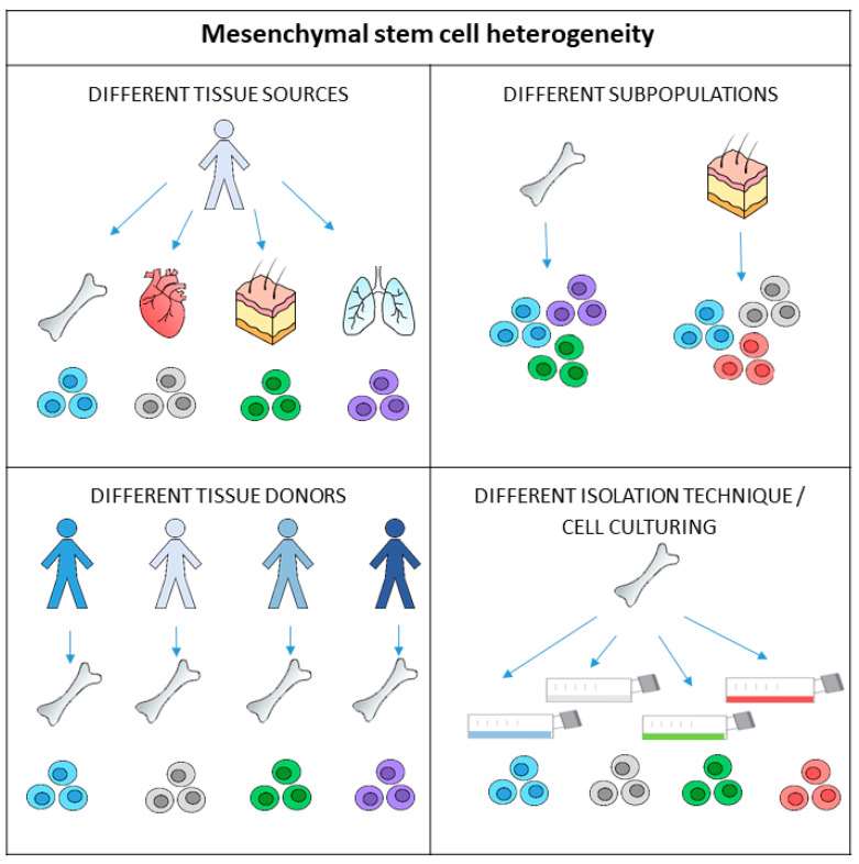 Fig. 1 MSCs exhibit heterogeneity on multiple levels.
Mesenchymal Stem Cells Counts per Total Nucleated Cells
MSC estimation per total nucleated cells is the simplest method for determining native MSC numbers, but it is inaccurate. Hernigou reported an equation for the prediction of the number of nucleated cells in bone marrow, as follows: N(108/kg) = (V × NP) - (V - 100) × NS/P, where V is the total volume of aspirate, NP is the nuclear cell count per ml of bone marrow aspirate, NS is the nuclear cell count per ml of peripheral blood, and P is the patient's weight. The above calculation was applied to MSCs isolated from bone marrow. Stem and progenitor cells in the uncultured stromal vascular fraction (SVF) from adipose tissue usually amount to up to 3% of the whole number of cells, which is 2500-fold greater than the frequency of stem cells in the bone marrow.
Estimation of Mesenchymal Stem Cell Count with the CFU-F Test
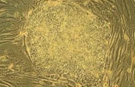
MSCs were initially referred to as fibroblast-like cells that can generate colony-forming unit fibroblasts or CFU-F. Determining the colony-forming units present is the oldest way of estimating the actual stem cells in the suspension of different cells. A suspension of mixed cells is plated on plastic culture dishes at a low density. Culturing under specific conditions allows stem cells to adhere, proliferate, and form colonies. After the growth of visible colonies, they are counted using light microscopy. The number of colonies indicates the number of cells that were able to proliferate, as each colony is presumed to originate from a single stem cell.
The CFU-F assay has some shortcomings. Cell culturing takes several days before the identification of colonies; the method is time-consuming and inappropriate for routine use, the counting of colonies is subjective, not standardized; and the technique lacks accuracy. Even though this procedure gives us the number of functional, proliferating MSCs in a tissue, there is a need for further investigation to explore alternative approaches to assessing the quantity of MSCs.
Mesenchymal Stem Cell Quantification with Immunophenotyping
For accurate counting of MSCs, their identity has to be ascertained first. Identification is based on the characteristic protein expression profile of MSCs. The Mesenchymal and Tissue Stem Cell Committee of the International Society for Cellular Therapy (ISCT) defined an MSC phenotype that is used irrespective of tissue source. Expanded cells are determined to be MSCs when expressing CD105, CD90, and CD73 and not expressing CD45, CD34, CD14 (CD11b), CD79 (CD19), or human lymphocyte antigen-DR (HLA-DR). This method is usually not used in the case of freshly isolated MSCs. Besides this, in recent years, new markers with positive or negative expression and new marker combinations have been recognized, among them CD49a, CD106 (VCAM-1), CD140b, CD146, CD271 (LNGFR), MSCA-1, GD2, and STRO-1. Many of these markers were used to identify MSC subpopulations expressing different types of regulatory proteins that function in hematopoiesis, angiogenesis, neural activities, and immunity processes.
To quantify MSCs before culturing, it is necessary to identify the MSC (sub)population from the harvested cell suspension where MSCs are still suspended with other cells. MSCs from the same tissue are heterogeneous and may not share the same phenotypical markers. Additionally, there is a lack of consensus on the markers identifying or distinguishing MSCs derived from different tissues. The chosen phenotype markers may influence the count; for example, the CD45-/CD271+ MSCs correlate more with CFU-F numbers than the CD45-/CD73+/CD90+/CD105+ MSCs. Therefore, the identification of native MSCs is much more complicated than expanded MSCs. The biological capacity of MSCs (i.e., immunomodulatory capacity, differentiation potential to a specific cell type, and endogenous stem cell mobilizing capacity) of one tissue source may differ from others. Isolation or production of a specific subpopulation of MSCs for a particular medical indication should improve outcomes.
Despite the numerous markers for MSC identification at our disposal, no agreement exists over the correct composite marker panel for defining MSCs in freshly isolated and non-cultured populations. For this reason, a precise characterization of MSCs derived from different tissues and their properties relating to their therapeutic potential represents an essential requirement for the exploitation and development of optimal MSC-based therapies. Additional studies should be conducted to test the correlation between phenotype markers and CFU-F and other biological effects. Together with multicentric study data, the ISCT could recommend criteria for native MSC analysis.
Creative Bioarray Relevant Recommendations
| Product/Service Types | Description |
| Mesenchymal Stem Cells | Creative Bioarray offers a broad range of mesenchymal stem cell products including cryopreserved human & animal MSCs from various tissue sources, optimized media for expansion and cryopreservation, kits and media to differentiate the cells into adipocytes and osteocytes, and characterization antibodies and kits. |
| MSC Validation Kits | Creative Bioarray's MSC validation kit provides a quantitative solution for the unambiguous identification of MSC. |
| Mesenchymal Stem Cells (MSCs) Services | Creative Bioarray provides advanced and experienced cell therapy research services covering all stages of MSC development, including isolation, expansion, characterization, and differentiation. |
RELATED PRODUCTS & SERVICES

Reference
- Maličev E, Jazbec K. (2024). "An Overview of Mesenchymal Stem Cell Heterogeneity and Concentration." Pharmaceuticals (Basel). 17 (3): 350.
