How to Assess the Migratory and Invasive Capacity of Cells?
Understanding the mechanisms underlying cell migratory and invasive capacity is crucial for several reasons. In developmental biology, cell migration is essential for processes such as gastrulation, neural crest cell migration, and organogenesis. In wound healing, the migration of cells is necessary for the re-epithelialization and restoration of tissue integrity. In the immune system, cell migration allows immune cells to move to sites of infection or inflammation. In cancer, the ability of tumor cells to migrate and invade surrounding tissues is a hallmark of metastasis, the primary cause of cancer-related deaths.
Introduction of Cell Migratory and Invasive Capacity
Cell migration is a complex and highly regulated process that involves coordinated changes in cell shape, adhesion, cytoskeletal dynamics, and signaling pathways. Cells can migrate individually or collectively, depending on the context and cellular interactions. In individual cell migration, cells extend protrusions at the leading edge, adhere to the extracellular matrix or neighboring cells, and generate contractile forces to propel themselves forward. Collective cell migration involves groups of cells moving in a coordinated manner, maintaining cell-cell contacts and communication.
Cell invasion, on the other hand, refers to the ability of cells to penetrate through barriers, such as the extracellular matrix, basement membranes, or other cellular layers. Invasion is a critical step in various biological processes, including immune cell infiltration during inflammation and the spread of cancer cells during metastasis. Invasive cells often acquire specialized properties, such as enhanced proteolytic activity to degrade surrounding tissues and increased motility to navigate through complex environments.
Approaches to Assess Cell Migratory and Invasive Capacity
- Scratch/wound healing assay. The scratch or wound healing assay is a widely used and straightforward method to evaluate cell migration. In this assay, cells are grown to confluence in a monolayer, and a uniform scratch is created using a sterile pipette tip or a specialized tool. The closure of the scratch over time indicates the migratory capacity of the cells. This assay is suitable for studying the migration of various cell types and can be easily adapted for high-throughput screening.
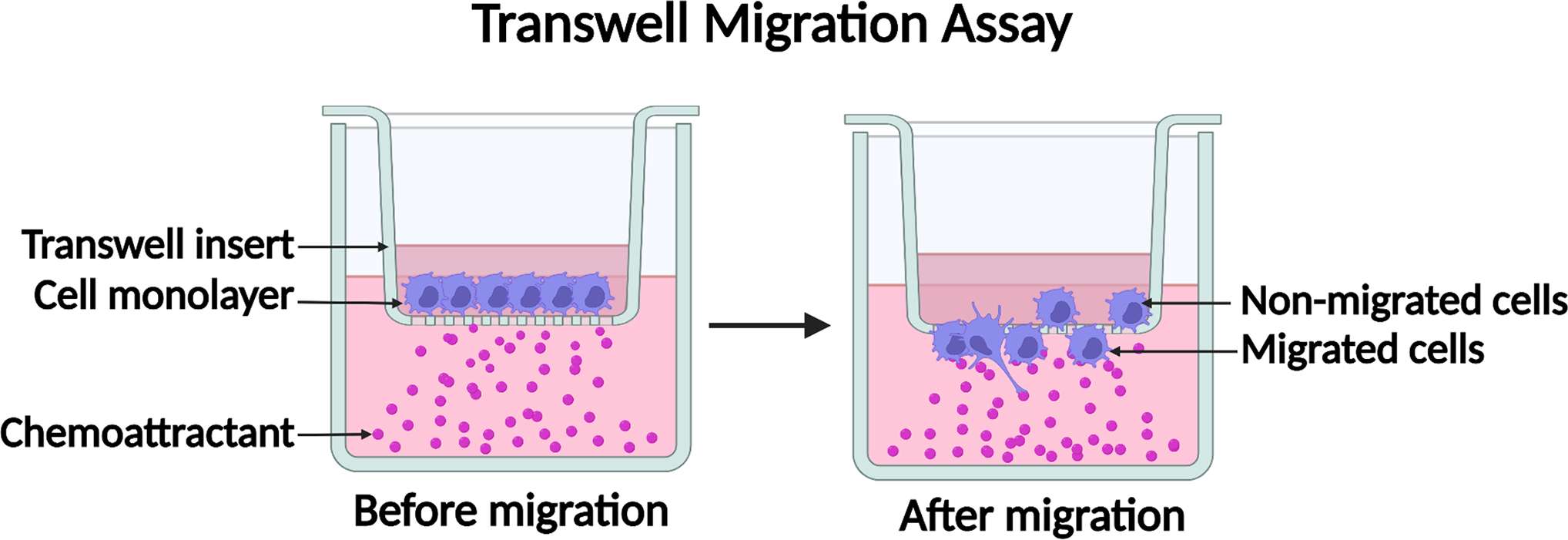
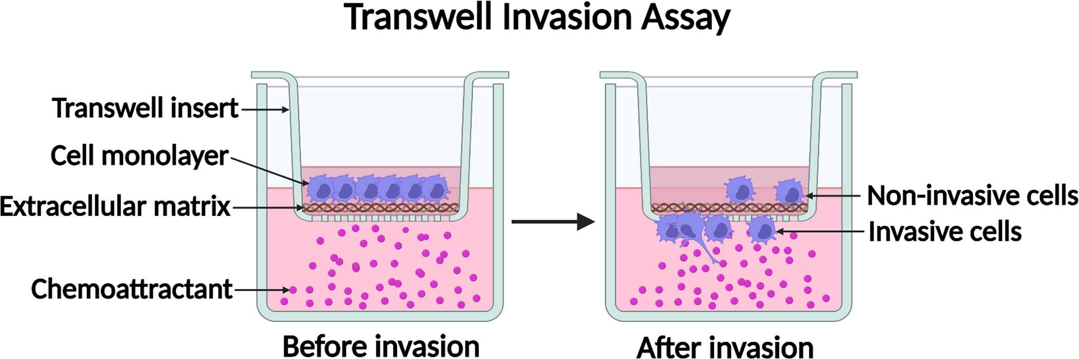
- Transwell assay/Boyden chamber assay. The transwell or Boyden chamber assay is another commonly employed technique to assess both cell migration and invasion. The assay utilizes a porous membrane insert placed in a multiwell plate. Cells are seeded in the upper chamber, while the lower chamber contains a chemoattractant that promotes cell migration. The migration of cells through the membrane is quantified by staining and counting the cells on the lower surface. To assess invasive capacity, the membrane can be pre-coated with extracellular matrix components such as Matrigel. This assay provides a quantitative measurement of cell migration and invasion and is suitable for studying chemotaxis.
 Fig.1 Diagram of the transwell cell migration assay. (Justus CR, et al., 2023)
 Fig.2 Diagram of the transwell cell invasion assay. (Justus CR, et al., 2023)
- Cell exclusion zone assay. The cell exclusion zone assay, also known as the "gap closure" assay, is similar to the scratch assay but offers a more precise and quantitative assessment of cell migration. In this method, a culture insert is placed in a cell culture dish to create a defined cell-free zone. After removing the insert, cells migrate into the zone, and the closure of the gap is measured and analyzed. This assay is particularly useful for studying the effect of various treatments or factors on cell migration.
- Fence assay (ring assay). The fence assay, or ring assay, is a specialized technique that enables the assessment of collective cell migration. In this assay, cells are plated within a circular pattern created by a removable ring or stencil. As the cells migrate, they maintain their cohesive nature and move collectively, allowing the study of coordinated migration. This assay is valuable for investigating processes such as epithelial sheet migration during embryogenesis or wound healing.
- Microcarrier bead assay. The microcarrier bead assay is a three-dimensional (3D) culture-based method that evaluates cell migration in a more physiologically relevant context. Cells are cultured on microcarrier beads, which provide a 3D environment mimicking the extracellular matrix. The migration of cells from one bead to another can be monitored and quantified, enabling the study of cell migration behavior in a more complex environment. This assay is particularly useful for studying the migration of tumor cells and assessing their response to therapeutic agents.
- Spheroid migration. Spheroid migration assays involve the use of multicellular spheroids as a model for studying cell migration and invasion. Spheroids, formed by aggregating cells in a non-adherent environment, mimic the structural and spatial complexity of tumors. Various methods, such as the hanging drop method or the liquid overlay technique, can be employed to generate spheroids. The migration of cells from the spheroid can be monitored and quantified, providing insights into their invasive potential and response to anti-migratory treatments.
- Capillary chamber migration assays (microfluidic chamber assays). Capillary chamber migration assays, also known as microfluidic chamber assays, utilize microfabrication techniques to create controlled microenvironments for cell migration studies. These devices consist of microchannels or chambers through which cells can migrate in response to chemical gradients or physical cues. The precise control over the microenvironment allows the investigation of cell migration behavior under more defined conditions, such as shear stress or concentration gradients. Microfluidic chamber assays offer high reproducibility and enable the study of cell migration in a tissue-like context.
Selection of Proper Methods for Cell Migration and Invasion
The choice of the appropriate method for assessing cell migration and invasion depends on several factors, including the research objectives, cell type, experimental conditions, and desired level of quantification. Each assay has its advantages and limitations, and researchers should carefully consider these factors before selecting a method.
- For instance, scratch and wound healing assays are simple and cost-effective but may not provide detailed information about invasive capacity. Transwell and Boyden chamber assays offer quantitative measurements and the ability to study invasion but may not fully capture the complexity of 3D environments. The cell exclusion zone assay provides a precise measurement of migration but may require specialized equipment. The fence assay is suitable for studying collective migration but may not apply to all cell types.
- If researchers aim to study cell migration in a more physiologically relevant context, 3D culture-based assays such as the microcarrier bead assay or spheroid migration assays can be employed. These assays allow cells to migrate in a 3D environment, providing insights into their behavior in complex tissue-like settings.
- For precise control over the microenvironment and the ability to study migration under defined conditions, capillary chamber migration assays or microfluidic chamber assays offer advantages. These assays enable the investigation of migration in response to specific chemical gradients or physical cues, providing a more detailed understanding of cell behavior.
Creative Bioarray Relevant Recommendations
| Product/Service Types | Description |
| Cell Migration and Invasion Assay Services | With a comprehensive list of cell lines in our inventory, Creative Bioarray provides cell migration and invasion assay services to all of our customers. |
| Cell Line Testing and Assays | Creative Bioarray has performed thousands of cell line testing services for clients in biopharma under cGMP and validated to current ICH guidelines. |
| Human Tumor Cells | Creative Bioarray provides human tumor cells sourced from a variety of tissue types. |
| Animal Tumor Cells | Creative Bioarray's animal tumor cells cover dozens of different animal species. We have hundreds of animal tumor cells as well as normal tissue cells from various organs and tissues. |
Reference
- Justus CR, et al. (2023). "Transwell In Vitro Cell Migration and Invasion Assays." Methods Mol Biol. 2644: 349-359.

