Exosome MicroRNA-22 Inhibiting Proliferation, Migration, and Invasion in OS
Scientific Reports. 2024 Jan 8; 14 (1): 761
Authors: Ruan Q, Wang C, Wu Y, Zhu Q
INTRODUCTION
Osteosarcoma (OS) is a primary malignant tumor that tends to occur in adolescents. The most common symptom is local pain at the epiphyseal end of the long shaft. The incidence rate of OS in adolescents is 8-11/million/year. MicroRNAs (miRNA) are the main cargo in exo-MSC, which participated in many diseases including OS. miR-22 is an evolutionarily conservative gene located on chromosome 17p13. The promoter transcription start site lacks a TATA box. miR-22 is abnormally up-regulated or down-regulated in many cancers, such as prostate cancer, esophageal squamous cell carcinoma, breast cancer, and gastric cancer.
METHODS
- MSC, MG63, and Saos were cultured in DMEM in 10% fetal bovine serum at 37°C in a humidified 5% CO2 incubator. The medium was replaced every 1-2 days. Exosomes (50 ug/ml) were added into the culture medium for co-culture experiments. miR-22 mimics and pcDNA3.1-Twist1 vector were used to overexpression of miR-22 and Twist1, respectively. miR-22 inhibitor and Twist1 siRNA (si-Twist1) were used to downexpression of miR-22 and Twist1, respectively.
- The proliferation of MG63 transfected with miR-22 mimics or inhibitor, Twist1 vector, or Twist1 siRNA was determined by the Cell Counting Kit-8 (CCK-8) assay, colony formation assay, and EdU assay according to the manufacturer's instructions. The migration and invasion of MG63 transfected with miR-22 mimics or inhibitors, Twist1 vector, or Twist1 siRNA were determined by transwell assay and wound healing assay.
- Culture medium was centrifuged at 300 g for 10 min, 2000 g for 20 min, 10,000 g for 30 min, and 100,000 g for 90 min, re-suspended and centrifuged at 100,000 g for 90 min. Exosomes were fixed in 2% glutaraldehyde and postfixed in 1% osmium tetroxide. Ultrathin sections were cut and stained with 10% uranyl acetate and 1% lead citrate. The ultrastructure of the exosome was observed by using a transmission electron microscope, Exosomes derived from MSCs were labeled with lipophilic dye PKH26 liquor.
- Browse our recommendations
| Product/Service Types | Description |
| Human Tumor Cells | Creative Bioarray provides human tumor cells sourced from a variety of tissue types. Our human tumor cells have the original pathological diagnosis and are analyzed for key mutations, presenting the real characteristics of their in vivo state and maintaining heterogeneity across multiple passages. |
| Cell Proliferation Assay Services | Creative Bioarray provides cell proliferation assay services for our customers. We provide optional assay methods based on the cell type and protocol and the customer's preference for proliferation measurement. |
| Cell Migration and Invasion Assay Services | With a comprehensive list of cell lines in our inventory, Creative Bioarray provides cell migration and Invasion Assay Services to all of our customers. |
| Exosome Isolation and Purification | Creative Bioarray provides reliable techniques for exosome isolation from different sample matrices. We also provide exosome isolation using specified techniques if required by customers |
| Exosome Quantification | Exosome quantification mainly depends on their characteristic physical properties, such as size, mass, and density, or membrane proteins present on their surface. Creative Bioarray offers a range of options to meet most exosome quantitation demands. |
RESULTS
- To verify the previous finding that miR-22 participated in the regulation of OS pathogenesis, miR-22 mimic and inhibitor were transfected into MG63. PCR and western blotting confirmed the transfected efficiency. High expression of miR-22 was found in MG63 transfected with miR-22 mimic, down expression of Twist1 and high expression of CADM1 were found in MG63 transfected with miR-22 mimic. MTS assay, CCK-8 assay, and colony formation assay showed upregulated proliferative of MG63 with low expression of miR-22 and downregulated proliferative of MG63 with high expression of miR-22. RT-qPCR and western blotting showed high expression of PCNA in MG63 with low expression of miR-22. miR-22 mimics reduced cell proliferation, as confirmed by EdU proliferation assays. Transwell assay and wound healing assay showed downregulated migration and invasion of MG63 with high expression of miR-22. RT-qPCR and western blotting showed up-regulated expression of MMP2 in MG63 with low expression of miR-22. These results showed that miR-22 inhibits MG63 proliferative, migration, and invasion.
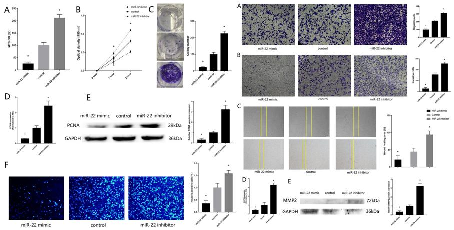 Fig. 1 Left: Downregulation of miR-22 regulates MG63 proliferative; Right: Downregulation of miR-22 regulates MG63 migration and invasion.
- The transfection efficiency of Twist1 vector, si-Twist1, miR-22 mimic and inhibitor were assessed by RT-qPCR. The rescue experiment showed that MG63 transfected with miR-22 mimic and Twist1 vector could remedy the reduced proliferation, migration, and invasion effectively caused by MG63 transfected with miR-22 mimic only. Low expression of Twist1 in MG63 could remedy the raised proliferation, migration, and invasion effect caused by MG63 transfected with miR-22 inhibitor only. Twist1 vector could also remedy the effect of the EMT signal pathway caused by MG63 transfected with miR-22 mimic only. A dual luciferase reporter assay was performed to confirm the binding sites between miR-22 and Twist1. miR-22 overexpression in MG63 cells reduced luciferase activity in cells expressing wild-type Twist1 compared with that in MG63 cells transfected with the mimic control and mutant Twist1. These results showed that miR-22 inhibits MG63 proliferative, migration and invasion through binding Twist1.
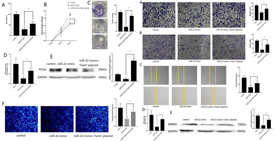 Fig. 2 Left: miR-22/Twist1 regulates MG63 proliferation; Right: miR-22/Twist1 regulates MG63 migration and invasion.
- To verify the effect of miR-22 cargo in exo-MSC inhibit MG63 proliferative, migration, and invasion, miR-22 mimic and inhibitor were transfected in MSC, which acquire high expression exosomes original MSC (high-miR22-exo-MSC) and low expression exosomes original MSC (low-miR22-exo-MSC). The transfect efficiency was confirmed by RT-qPCR. Then the modified exosomes were added to the MG63. Low proliferative, migration, and invasion ability were found in MG63 co-culture with high-miR22-exo-MSC, and High proliferative, migration, and invasion ability were found in MG63 co-culture with low-miR22-exo-MSC. The functions of miR-22 cargo in exo-MSC in Saos were also verified.
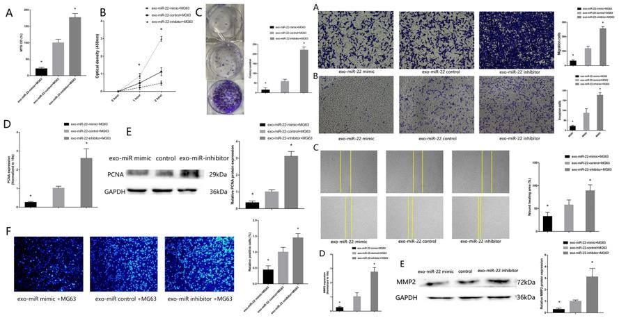 Fig. 3 Left: Exosome miR-22 original MSC inhibit MG63 proliferative; Right: Exosome miR-22 original MSC inhibit MG63 migration and invasion.
SUMMARY
This study aims to the function of miR-22 original mesenchymal stem cells (MSC) on OS proliferation, migration, and invasion. MTS assay, CCK8 assay, colony forming assay, and EdU assay were performed to detect the proliferation effect of miR-22 on MG63. Transwell migration assay, transwell invasion assay, and wound healing assay were used to verify the migration and invasion effect of miR-22 on MG63. Luciferase reporter assay confirms the binding sites between miR-22 and Twist1. RT-qPCR confirmed miR-22 and CADM1 downregulated and Twist1 upregulated in OS tissues, MG63 and Saos. Exosome original MSC labeled with PKH-26 could be uptake by MG63, which upregulated the expression of miR-22 in MG63. High expression of miR-22 in MG63 inhibited proliferation, migration, and invasion, which could be rescued by Twist1. Dual luciferase reporter analysis confirmed Twist1 was a target of miR-22. Exosomes modified with miR-22 mimic inhibit proliferation, migration, and invasion more efficiently than exosome original MSC. miR-22 cargo in exo-MSC could uptake by MG63 and supply MG63 with miR-22, which inhibit MG63 proliferation, migration and invasion through targeting Twist1.
RELATED PRODUCTS & SERVICES


Reference
- Ruan Q, et al. (2024). "Exosome microRNA-22 inhibiting proliferation, migration and invasion through regulating Twist1/CADM1 axis in osteosarcoma." Sci Rep. 14 (1): 761.