- You are here: Home
- Disease Models
- Inflammation & Autoimmune Disease Models
- Multiple Sclerosis (MS) Models
- EAE Models
- Myelin Oligodendrocyte Glycoprotein (MOG)-Induced Experimental Autoimmune Encephalomyelitis (EAE) Model
Disease Models
- Oncology Models
-
Inflammation & Autoimmune Disease Models
- Rheumatoid Arthritis Models
- Glomerulonephritis Models
- Multiple Sclerosis (MS) Models
- Ocular Inflammation Models
- Sjögren's Syndrome Model
- LPS-induced Acute Lung Injury Model
- Peritonitis Models
- Passive Cutaneous Anaphylaxis Model
- Delayed-Type Hypersensitivity (DTH) Models
- Inflammatory Bowel Disease Models
- Systemic Lupus Erythematosus Animal Models
- Asthma Model
- Sepsis Model
- Psoriasis Model
- Atopic Dermatitis (AD) Model
- Scleroderma Model
- Gouty Arthritis Model
- Carrageenan-Induced Air Pouch Synovitis Model
- Carrageenan-Induced Paw Edema Model
- Experimental Autoimmune Myasthenia Gravis (EAMG) Model
-
Cardiovascular Disease Models
- Surgical Models
- Animal Models of Hypertension
- Venous Thrombosis Model
- Atherosclerosis model
- Cardiac Arrhythmia Model
- Hyperlipoidemia Model
- Doxorubicin-induced Heart Failure Model
- Isoproterenol-induced Heart Failure Model
- Arterial Thrombosis Model
- Pulmonary Arterial Hypertension (PAH) Models
- Heart Failure with Preserved Ejection Fraction (HFpEF) Model
-
Neurological Disease Models
- Alzheimer's Disease Modeling and Assays
- Seizure Models
- Parkinson's Disease Models
- Ischemic Stroke Models
- Acute Spinal Cord Injury (ASCI) Model
- Traumatic Brain Injury (TBI) Model
- Hypoxic-Ischemic Encephalopathy (HIE) Model
- Tourette Syndrome (TS) Model
- Amyotrophic Lateral Sclerosis (ALS) Model
- Huntington's Disease (HD) Model
- Intracerebral hemorrhage (ICH) Models
- Pain Models
- Metabolic Disease Models
- Liver Disease Models
- Rare Disease Models
- Respiratory Disease Models
- Digestive Disease Models
-
Urology Disease Models
- Cisplatin-induced Nephrotoxicity Model
- Unilateral Ureteral Obstruction Model
- 5/6 Nephrectomy Model
- Renal Ischemia-Reperfusion Injury (RIRI) Model
- Diabetic Nephropathy (DN) Models
- Passive Heymann Nephritis (PHN) Model
- Adenine-Induced Chronic Kidney Disease (CKD) Model
- Kidney Stone Model
- Doxorubicin-Induced Nephropathy Model
- Orthopedic Disease Models
- Ocular Disease Models
- Skin Disease Models
- Infectious Disease Models
Myelin Oligodendrocyte Glycoprotein (MOG)-Induced Experimental Autoimmune Encephalomyelitis (EAE) Model
Creative Bioarray is proud to present the Myelin Oligodendrocyte Glycoprotein (MOG)-induced Experimental Autoimmune Encephalomyelitis (EAE) model, designed to facilitate our clients' investigations into multiple sclerosis (MS) treatments. This meticulously developed model offers a robust and reproducible environment for evaluating the efficacy of novel therapeutic agents. By providing our clients with this state-of-the-art resource, we ensure that they are equipped with the most advanced tools necessary for their MS research initiatives, enabling them to explore the potential of new compounds with confidence and precision.
Our Myelin Oligodendrocyte Glycoprotein (MOG)-Induced Experimental Autoimmune Encephalomyelitis (EAE) Model
- Model Description
The MOG-induced EAE model is a pivotal animal model used to study MS. MOG, a member of the immunoglobulin superfamily, is specifically expressed in the CNS on the surface of myelin sheaths and oligodendrocyte processes. This model is induced in female C57BL/6 mice by immunizing them with MOG peptide, followed by pertussis toxin (PTX) injections. The MOG peptide stimulates the proliferation and differentiation of MOG-specific autoimmune T cells, while PTX enhances disease progression by modulating the immune response and aiding the migration of these T cells into the CNS. This model closely mimics the neuroinflammation and demyelination observed in MS, making it an invaluable tool for developing MS therapeutics and studying the immune-mediated mechanisms of the disease.
Characteristics of the MOG-Induced EAE Model in Creative Bioarray
- Characterized by mononuclear inflammatory infiltration, including macrophages and CD4+ T cells.
- MOG-induced EAE model is considered the gold standard model and is routinely used in the development of therapeutics for MS.
- MOG-induced EAE model is a good model of chronic inflammatory demyelination.
- Available Animal
C57BL/6 mouse (female)
- Modeling Method
All animals, with the exception of those in the control group, are immunized to induce EAE by receiving a MOG35-55 emulsion through subcutaneous (s.c.) injection on Day 0, accompanied by PTX administered intraperitoneally (i.p.) on both Day 0 and Day 2.
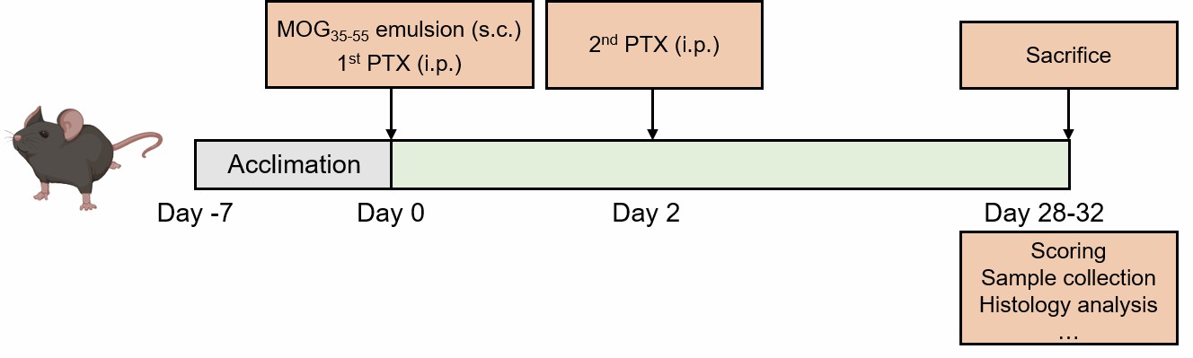 Fig. 1 Schematic diagram of the modeling method for MOG-induced EAE model.
- Endpoints
- Body weight
- Clinical score
- Histology analysis
- Cytokine analysis
- qPCR or Western blot
- Other customized endpoints
Example Data
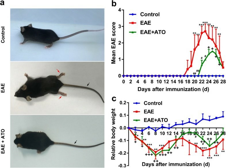 Fig. 2 ATO ameliorates EAE progression in mice. a Representative images show behavioral symptoms of EAE mice in individual groups. Black arrow presents limp tail, and red arrow presents hind limb paralysis. The clinical scores (b) and changes in body weight (c) were recorded. n = 10 mice/group. (An et al. 2020)
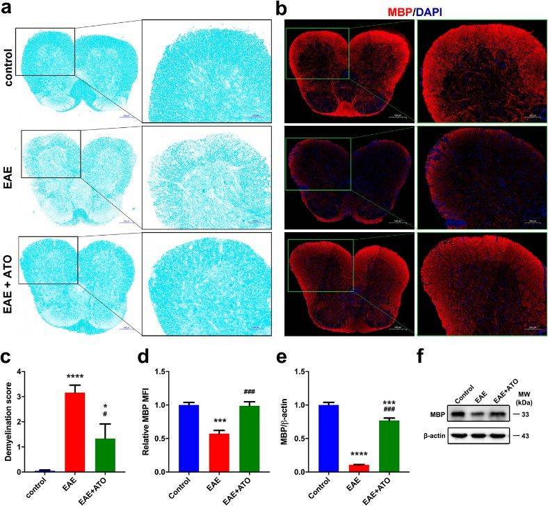 Fig. 3 ATO alleviates demyelination in the spinal cord of EAE mice. Representative LFB staining sections (a) and demyelination score (c) of spinal cord. b Representative immunofluorescent staining sections of MBP in spinal cord.. d Mean fluorescence intensity analysis of MBP in b. e The mRNA level of MBP in spinal cord. f The protein level of MBP in spinal cord. (An et al. 2020)
Furthermore, we also provide other EAE models that maybe you are interested in:
- Proteolipid Protein (PLP)-Induced Experimental Autoimmune Encephalomyelitis (EAE) Model
- Myelin Basic Protein (MBP)-Induced Experimental Autoimmune Encephalomyelitis (EAE) Model
Quotation and Ordering
Creative Bioarray is proud to offer a suite of comprehensive and tailored services for every project, made possible by our team of highly skilled scientists with a track record of excellence in pharmacology and efficacy studies. We are committed to delivering these services at the most competitive prices, ensuring that our clients receive exceptional value without compromising on quality. If you are interested in our services, please feel free to contact us at any time or submit an inquiry to us directly.
References
- Glatigny, S., Bettelli, E. Experimental autoimmune encephalomyelitis (EAE) as animal models of multiple sclerosis (MS). Cold Spring Harbor perspectives in medicine, 2018, 8(11): a028977.
- An, K., et al. Arsenic trioxide ameliorates experimental autoimmune encephalomyelitis in C57BL/6 mice by inducing CD4+ T cell apoptosis. Journal of Neuroinflammation, 2020, 17: 1-14.
For research use only. Not for any other purpose.
