- You are here: Home
- Services
- Disease Models
- Metabolic Disease Models
- Nonalcoholic Fatty Liver Disease Model
- High-Fat Diet-Induced Nonalcoholic Fatty Liver Disease (NAFLD) Model
Services
-
Cell Services
- Cell Line Authentication
- Cell Surface Marker Validation Service
-
Cell Line Testing and Assays
- Toxicology Assay
- Drug-Resistant Cell Models
- Cell Viability Assays
- Cell Proliferation Assays
- Cell Migration Assays
- Soft Agar Colony Formation Assay Service
- SRB Assay
- Cell Apoptosis Assays
- Cell Cycle Assays
- Cell Angiogenesis Assays
- DNA/RNA Extraction
- Custom Cell & Tissue Lysate Service
- Cellular Phosphorylation Assays
- Stability Testing
- Sterility Testing
- Endotoxin Detection and Removal
- Phagocytosis Assays
- Cell-Based Screening and Profiling Services
- 3D-Based Services
- Custom Cell Services
- Cell-based LNP Evaluation
-
Stem Cell Research
- iPSC Generation
- iPSC Characterization
-
iPSC Differentiation
- Neural Stem Cells Differentiation Service from iPSC
- Astrocyte Differentiation Service from iPSC
- Retinal Pigment Epithelium (RPE) Differentiation Service from iPSC
- Cardiomyocyte Differentiation Service from iPSC
- T Cell, NK Cell Differentiation Service from iPSC
- Hepatocyte Differentiation Service from iPSC
- Beta Cell Differentiation Service from iPSC
- Brain Organoid Differentiation Service from iPSC
- Cardiac Organoid Differentiation Service from iPSC
- Kidney Organoid Differentiation Service from iPSC
- GABAnergic Neuron Differentiation Service from iPSC
- Undifferentiated iPSC Detection
- iPSC Gene Editing
- iPSC Expanding Service
- MSC Services
- Stem Cell Assay Development and Screening
- Cell Immortalization
-
ISH/FISH Services
- In Situ Hybridization (ISH) & RNAscope Service
- Fluorescent In Situ Hybridization
- FISH Probe Design, Synthesis and Testing Service
-
FISH Applications
- Multicolor FISH (M-FISH) Analysis
- Chromosome Analysis of ES and iPS Cells
- RNA FISH in Plant Service
- Mouse Model and PDX Analysis (FISH)
- Cell Transplantation Analysis (FISH)
- In Situ Detection of CAR-T Cells & Oncolytic Viruses
- CAR-T/CAR-NK Target Assessment Service (ISH)
- ImmunoFISH Analysis (FISH+IHC)
- Splice Variant Analysis (FISH)
- Telomere Length Analysis (Q-FISH)
- Telomere Length Analysis (qPCR assay)
- FISH Analysis of Microorganisms
- Neoplasms FISH Analysis
- CARD-FISH for Environmental Microorganisms (FISH)
- FISH Quality Control Services
- QuantiGene Plex Assay
- Circulating Tumor Cell (CTC) FISH
- mtRNA Analysis (FISH)
- In Situ Detection of Chemokines/Cytokines
- In Situ Detection of Virus
- Transgene Mapping (FISH)
- Transgene Mapping (Locus Amplification & Sequencing)
- Stable Cell Line Genetic Stability Testing
- Genetic Stability Testing (Locus Amplification & Sequencing + ddPCR)
- Clonality Analysis Service (FISH)
- Karyotyping (G-banded) Service
- Animal Chromosome Analysis (G-banded) Service
- I-FISH Service
- AAV Biodistribution Analysis (RNA ISH)
- Molecular Karyotyping (aCGH)
- Droplet Digital PCR (ddPCR) Service
- Digital ISH Image Quantification and Statistical Analysis
- SCE (Sister Chromatid Exchange) Analysis
- Biosample Services
- Histology Services
- Exosome Research Services
- In Vitro DMPK Services
-
In Vivo DMPK Services
- Pharmacokinetic and Toxicokinetic
- PK/PD Biomarker Analysis
- Bioavailability and Bioequivalence
- Bioanalytical Package
- Metabolite Profiling and Identification
- In Vivo Toxicity Study
- Mass Balance, Excretion and Expired Air Collection
- Administration Routes and Biofluid Sampling
- Quantitative Tissue Distribution
- Target Tissue Exposure
- In Vivo Blood-Brain-Barrier Assay
- Drug Toxicity Services
High-Fat Diet-Induced Nonalcoholic Fatty Liver Disease (NAFLD) Model
Creative Bioarray is a leading and reliable industry leader specializing in the meticulous development of animal models that accurately replicate human diseases. Among our extensive repertoire of models, we have successfully created the high-fat diet-induced nonalcoholic fatty liver disease (NAFLD) model. This model is designed to facilitate a deeper understanding of NAFLD's pathogenesis and to evaluate potential drug candidates.
The high-fat diet -induced NAFLD model is a pivotal tool in our preclinical research arsenal, designed to closely mimic the pathogenesis of human NAFLD. This model utilizes diets rich in fat to induce obesity and NAFLD in rodents. The model's efficacy in replicating metabolic syndrome, hepatic steatosis, and nonalcoholic steatohepatitis (NASH) has been well-validated, with variations in diet composition and duration enabling the study of different aspects of the disease.
Our High-fat Diet-Induced Nonalcoholic Fatty Liver Disease (NAFLD) Model
- Available Animal
- Rat
- Mouse
- Modeling Method
Animals are fed a high-fat diet for 16-20 weeks to induce NAFLD.
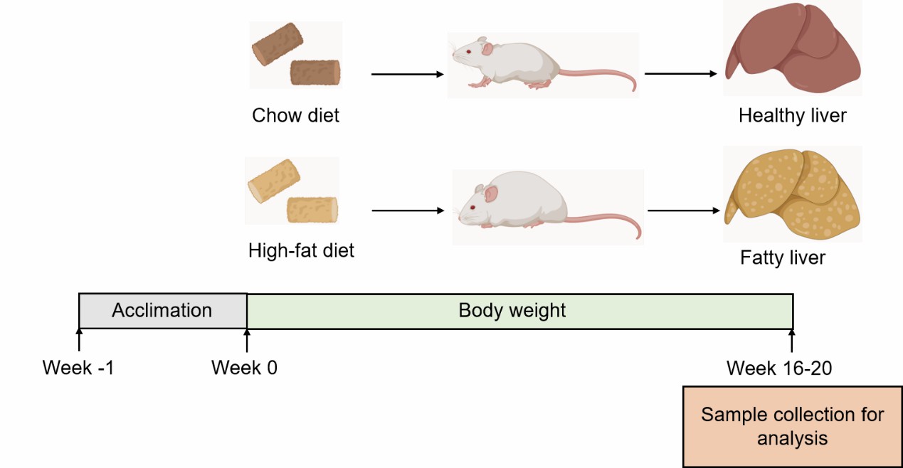 Fig. 1 Modeling method for the high-fat diet-induced NAFLD model at Creative Bioarray.
- Endpoints
- Body weight
- Liver weight
- Serum analysis: ALT, AST, TG, TC
- Histology analysis (liver): Oil red O staining
- Other customized endpoints
Example Data
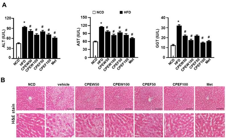 Fig. 2 Citrus peel extract protects against HFD-induced hepatic functional damage. (A) AST, ALT, and GGT were measured in HFD-fed rats. (B) H&E staining assay was performed using the liver. (Lee et al. 2020)
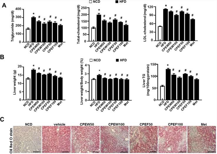 Fig 3 Citrus peel prevents HFD-induced hepatic steatosis. (A) Biochemical analysis of plasma samples. Levels of TG, TC and LDL-c were measured in the plasma of rat in different experimental groups. (B) Liver weight, and liver weight/body weight and liver TG content were measured 8 weeks after initial CPEW and CPEF administration. (C) Liver tissues were subjected to Oil Red O staining. (Lee et al. 2020)
Quotation and Ordering
Creative Bioarray is equipped with state-of-the-art preclinical technology platforms and backed by experienced research and scientific teams. Our comprehensive range of services is delivered with an unwavering commitment to high quality and efficiency, ensuring consistent exceptional high-quality results and rapid turnaround times. If you are interested in our services, please do not hesitate to contact us at any time or submit an inquiry to us directly.
References
- Fang, T., et al. Mouse models of nonalcoholic fatty liver disease (NAFLD): pathomechanisms and pharmacotherapies. International Journal of Biological Sciences, 2022, 18(15): 5681.
- Nevzorova, Y.A., et al. Animal models for liver disease–a practical approach for translational research. Journal of hepatology, 2020, 73(2): 423-440.
- Lee G H, Peng C, Park S A, et al. Citrus peel extract ameliorates high-fat diet-induced NAFLD via activation of AMPK signaling[J]. Nutrients, 2020, 12(3): 673.
Explore Other Options
For research use only. Not for any other purpose.
