- You are here: Home
- Disease Models
- Neurological Disease Models
- Intracerebral hemorrhage (ICH) Models
- Collagenase-Induced Intracerebral Hemorrhage (ICH) Model
Disease Models
- Oncology Models
-
Inflammation & Autoimmune Disease Models
- Rheumatoid Arthritis Models
- Glomerulonephritis Models
- Multiple Sclerosis (MS) Models
- Ocular Inflammation Models
- Sjögren's Syndrome Model
- LPS-induced Acute Lung Injury Model
- Peritonitis Models
- Passive Cutaneous Anaphylaxis Model
- Delayed-Type Hypersensitivity (DTH) Models
- Inflammatory Bowel Disease Models
- Systemic Lupus Erythematosus Animal Models
- Asthma Model
- Sepsis Model
- Psoriasis Model
- Atopic Dermatitis (AD) Model
- Scleroderma Model
- Gouty Arthritis Model
- Carrageenan-Induced Air Pouch Synovitis Model
- Carrageenan-Induced Paw Edema Model
- Experimental Autoimmune Myasthenia Gravis (EAMG) Model
-
Cardiovascular Disease Models
- Surgical Models
- Animal Models of Hypertension
- Venous Thrombosis Model
- Atherosclerosis model
- Cardiac Arrhythmia Model
- Hyperlipoidemia Model
- Doxorubicin-induced Heart Failure Model
- Isoproterenol-induced Heart Failure Model
- Arterial Thrombosis Model
- Pulmonary Arterial Hypertension (PAH) Models
- Heart Failure with Preserved Ejection Fraction (HFpEF) Model
-
Neurological Disease Models
- Alzheimer's Disease Modeling and Assays
- Seizure Models
- Parkinson's Disease Models
- Ischemic Stroke Models
- Acute Spinal Cord Injury (ASCI) Model
- Traumatic Brain Injury (TBI) Model
- Hypoxic-Ischemic Encephalopathy (HIE) Model
- Tourette Syndrome (TS) Model
- Amyotrophic Lateral Sclerosis (ALS) Model
- Huntington's Disease (HD) Model
- Intracerebral hemorrhage (ICH) Models
- Pain Models
- Metabolic Disease Models
- Liver Disease Models
- Rare Disease Models
- Respiratory Disease Models
- Digestive Disease Models
-
Urology Disease Models
- Cisplatin-induced Nephrotoxicity Model
- Unilateral Ureteral Obstruction Model
- 5/6 Nephrectomy Model
- Renal Ischemia-Reperfusion Injury (RIRI) Model
- Diabetic Nephropathy (DN) Models
- Passive Heymann Nephritis (PHN) Model
- Adenine-Induced Chronic Kidney Disease (CKD) Model
- Kidney Stone Model
- Doxorubicin-Induced Nephropathy Model
- Orthopedic Disease Models
- Ocular Disease Models
- Skin Disease Models
- Infectious Disease Models
Collagenase-Induced Intracerebral Hemorrhage (ICH) Model
Creative Bioarray offers a meticulously characterized intracerebral hemorrhage (ICH) model, induced by collagenase, specifically designed for in-depth neuroprotection studies. Our comprehensive services extend to a wide array of behavioral assessments, functional recovery evaluations, and detailed neuropathological analyses, all of which can be tailored to meet the precise requirements of our clients.
Bacterial collagenase, a potent protease, targets and degrades the extracellular matrix surrounding brain capillaries, leading to their weakening and eventual rupture. This breach results in the extravasation of blood, mirroring the spontaneous intracerebral bleeding observed in approximately 30% of patients diagnosed with ICH. The innovative use of bacterial collagenase injection into the basal ganglia to induce the breakdown of the vascular basement membrane was pioneered in the early 1990s, initially employing mouse models. Since its inception, this technique has gained widespread adoption due to its ability to closely mimic the pathophysiological processes of ICH in humans. This model not only enhances our understanding of the disease mechanisms but also provides a valuable platform for the development and testing of potential therapeutic strategies aimed at mitigating the devastating effects of ICH.
Our Collagenase-Induced Intracerebral Hemorrhage (ICH) Model
- Available Animal
- Rat
- Mouse
- Modeling Method
The animals are first anesthetized and then securely positioned on a stereotaxic apparatus. A precise burr hole, measuring 0.5 mm in diameter, is drilled at specific coordinates in relation to bregma. Next, a solution of saline enriched with collagenase is carefully administered using a 1 μl microsyringe, at a controlled rate of 2 μl per minute. After the infusion, the needle is left in place for an additional 10 minutes before being withdrawn. In contrast, the sham group undergoes similar procedures without the infusion of collagenase.
- Endpoints
- Clinical score: Longa score, Bederson score
- Survival rate
- Histology analysis: H&E staining, TUNEL staining, IHC
- Cytokine analysis
- Brain water content
- qPCR or Western blot
- Other customized endpoints
Example Data
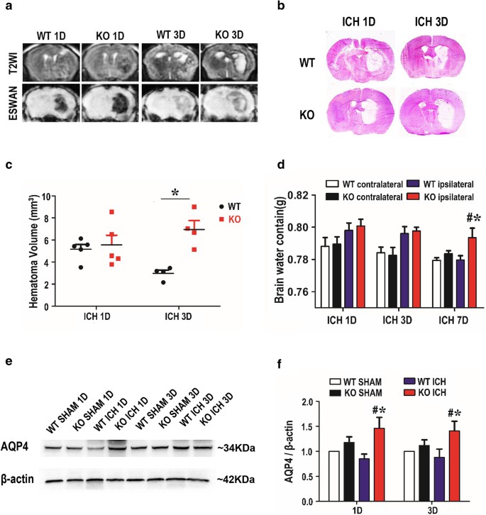 Fig. 1 TREK-1-deficient mice possess a larger hematoma volume on day 3 after ICH and more brain water content on day 7 post-ICH. a A representative MRI image on days 1 and 3 after ICH. b Representative image of HE staining on days 1 and 3 after ICH. c Statistic analysis of hematoma volume according to the HE staining. d Statistical analysis of brain water content using the wet/dry weigh method on days 1, 3, and 7 after ICH. e Representative western blot image of AQP4 expression in the perihematoma tissue. f Quantification of AQP4 expression according to the β-actin expression on days 1 and 3 after ICH. (Fang et al. 2019)
Meanwhile, we also provide another ICH model that maybe you are interested in:
Quotation and Ordering
Creative Bioarray establishes flexibly tailored study protocols that fit each therapeutics testing requirements. Whether it's adjusting the study design, modifying the selection of animal models, or refining the analytical methods, our approach ensures that each therapeutic is evaluated under the most appropriate and effective conditions. This tailored strategy not only accelerates the discovery process but also significantly contributes to the development of safer, more efficacious treatments. If you are interested in our services, please feel free to contact us at any time or submit an inquiry to us directly.
References
- Fang, Y., et al. Deficiency of TREK-1 potassium channel exacerbates blood-brain barrier damage and neuroinflammation after intracerebral hemorrhage in mice. Journal of neuroinflammation, 2019, 16: 1-16.
- Paiva, W.S., et al. Animal models for the study of intracranial hematomas (Review). Exp Ther Med, 2022; 25(1):20.
For research use only. Not for any other purpose.
