Featured Products
Our Promise to You
Guaranteed product quality, expert customer support

ONLINE INQUIRY
OPM-2
Cat.No.: CSC-C0222
Source: multiple myeloma
Morphology: single, round to polygonal cells in suspension
Culture Properties: suspension
- Specification
- Background
- Scientific Data
- Q & A
- Customer Review
Immunology: CD3 -, CD10 -, CD19 -, CD20 -, CD34 -, CD37 -, CD38 +, CD80 -, CD138 +, HLA-DR -, sm/cyIgG -, sm/cyIgM -, sm/cykappa -, sm/cylambda -
Viruses: ELISA: reverse transcriptase negat
The OPM-2 cell line was established in 1982 from the peripheral blood of a 56-year-old woman who had been diagnosed with multiple myeloma in the IgG lambda subtype. At the time of cell line derivation, the patient was in the leukemic phase of the disease, which was characterized as a relapse in the terminal stage.
The OPM-2 cells are notable for the presence of the t(4;14) chromosomal translocation. This cytogenetic abnormality leads to the formation of a fusion gene known as IGH-FGFR3, which is also referred to as IGH-FGFR3 or IGH-MMSET. The expression of this fusion protein is a defining molecular feature of the OPM-2 cell line and is closely linked to the pathogenesis of multiple myeloma in this patient.
Morphologically, the OPM-2 cells are described as single, round to polygonal cells that grow in suspension culture. As the cell line is of human origin, it is considered a valuable model for studying the biology and treatment of multiple myeloma, particularly in the context of the IGH-FGFR3 fusion gene and its role in driving disease progression. The availability of the OPM-2 cell line has been instrumental in advancing our scientific understanding of this hematological malignancy.
Increased Adhesion of OPM-2 Cells to HUVECs After Treatment With CSP
The molecular mechanism underlying the interaction between myeloma cells and stromal cells was investigated by using a human myeloma cell line (OPM-2) and human umbilical vein endothelial cells (HUVECs). Adhesion of OPM-2 cells to HUVECs was found to be significantly augmented with the treatment of OPM-2 cells with an α-glycosidase inhibitor, castanospermine (CSP).
OPM-2 cells were cultured with or without CSP (200 μg/mL) for 48 hours and then cocultured with HUVECs. In the absence of CSP treatment, 17.8% of OPM-2 cells adhered to HUVECs (Fig. 1). The preincubation of OPM-2 cells with CSP led to a significant increase in adhesion of OPM-2 cells, and 24.6% of OPM-2 cells were found to adhere to HUVECs (Fig. 1). The effect of CSP on surface expression of adhesion molecules was examined, which were known to express on myeloma cells. Flow cytometry analysis showed that CD44 and CD49d were expressed on OPM-2 cells at a high level, and CD54 at a moderate level, whereas little or no expression of CD11a, CD18, CD49e, or LAM-1 was observed on the cells (Table 1). The surface expression of the adhesion molecules was not influenced by the treatment of CSP (Table 1). These results indicated that CSP did not affect the expression of the well-known adhesion molecules and suggested that CSP might influence the function of adhesion molecules by altering their oligosaccharide structures.
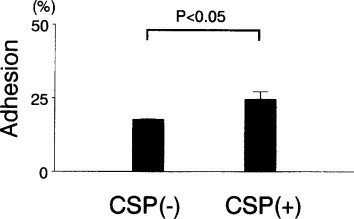 Fig. 1 Effect of CSP on the adhesion of OPM-2 cells to HUVECs. (Nishiura T, et al., 1996)
Table 1. Effect of CSP treatment on surface expression of adhesion molecules.
| Treatment | Proportions of Positive Cells (%) | ||||||
| CD11a | CD18 | CD44 | CD54 | CD49d | CD49e | LAM-1 | |
| Control | 0 | 0 | 93.7 | 31.1 | 96.8 | 0 | 4.6 |
| CSP | 0 | 0 | 95.5 | 28.7 | 96.6 | 0 | 4.2 |
MSCs Inhibit the Effects of Dexamethasone on Multiple Myeloma Cells
Mesenchymal stem cells (MSCs) participate in the occurrence and development of multiple myeloma. Multiple myeloma cells (OPM-2 and RPMI8226 cells) were cocultured with MSCs with or without dexamethasone. Treatment with dexamethasone (10 μM) significantly decreased the cell viability of OPM-2 and RPMI8226 cells (Fig. 2a and 2b). However, the presence of MSCs increased the cell viability of OPM-2 and RPMI8226 cells as compared to the dexamethasone-treated cells (Fig. 2a and 2b). Treatment with dexamethasone (10 μM) significantly decreased the cell number and colony number, respectively (Fig. 2c-f). Interestingly, the presence of MSCs increased the cell and colony numbers of OPM-2 and RPMI8226 cells as compared to the dexamethasone-treated cells (Fig. 2c-f).
Treatment with dexamethasone (10 μM) significantly increased the percentage of apoptotic OPM-2 and RPMI8226 cells (Fig. 3a and 3b). However, the presence of MSCs decreased the percentage of apoptotic OPM-2 and RPMI8226 cells as compared to the dexamethasone-treated cells (Fig. 3a and 3b). Moreover, cells were arrested at the G0/G1 phase in the dexamethasone (10 μM)-treated group (Fig. 3c and 3d). Interestingly, the presence of MSCs increased the percentage of cells in the S phase, while decreasing the percentage of cells in the G0/G1 phase.
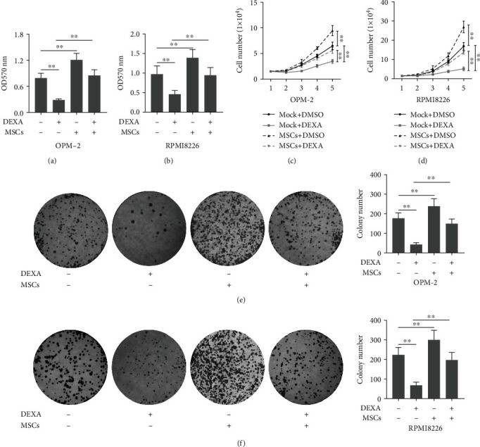 Fig. 2 The presence of MSCs suppressed the inhibitory effects of dexamethasone on cell proliferation. (Deng M, et al., 2022)
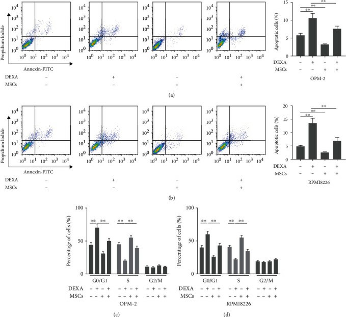 Fig. 3 The presence of MSCs regulated the effects of dexamethasone on cell apoptosis and cell cycle distribution. (Deng M, et al., 2022)
Cell-cycle control depends exclusively on post-transcriptional mechanisms that involve the regulation of Cdk activity by phosphorylation and the binding of regulatory proteins such as cyclins, which are themselves regulated by proteolysis.
The OPM-2 cell line is characterized by the presence of the t(4;14) translocation, which leads to the IGH@-FGFR3 (IGH-FGFR3; IGH-MMSET) fusion gene.
The OPM-2 cell line is of human origin, derived from a patient with multiple myeloma.
The OPM-2 cell line is considered a valuable model for studying the biology and treatment of multiple myeloma, particularly in the context of the IGH-FGFR3 fusion gene and its role in driving disease progression.
Ask a Question
Average Rating: 5.0 | 3 Scientist has reviewed this product
Impressive quality and speed
The tumor cell products arrived faster than expected and the quality is exceptional.
14 Aug 2022
Ease of use
After sales services
Value for money
High-quality products
The quality of the cells is outstanding and they have met all of our experimental requirements.
11 Mar 2024
Ease of use
After sales services
Value for money
Responsive customer services
The customer service team was very responsive, providing all necessary documentation and assistance promptly. I highly recommend Creative Bioarray for cell line purchases.
23 Apr 2024
Ease of use
After sales services
Value for money
Write your own review
- You May Also Need

