Featured Products
Our Promise to You
Guaranteed product quality, expert customer support

ONLINE INQUIRY
- Specification
- Background
- Scientific Data
- Q & A
- Customer Review
The human granulosa-like tumor cell line KGN was originally established in 2001 by Nishi and colleagues from a patient with invasive ovarian granulosa cell carcinoma. KGN cells have a slow doubling time (46.4 hours), which is typical of granulosa cell tumors, and can maintain a differentiated state in vitro.
Compared with the five human ovarian granulosa cell lines developed before 2001, KGN cells not only retain most of the physiological activities of granulosa cells, but also express the follicle-stimulating hormone (FSH) receptor, like primary granulosa cells do. Under the stimulation of FSH and human menopausal gonadotropin (HMG), aromatase activity increases, leading to the secretion of estrogen and other hormones. As a consequence, in addition to the study of their tumorigenic properties, KGN cells are often used as a model for granulosa cell function to study the mechanisms involved in follicular development, hormone regulation, granulosa cell tumors, and related diseases (such as polycystic ovary syndrome, PCOS).
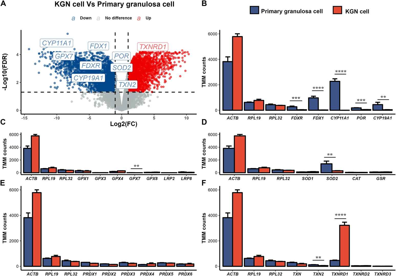 Fig. 1. Comparison of gene expression data between KGN and primary granulosa cells (Tang, Feng, Katja Hummitzsch, and Raymond J. Rodgers. 2024).
O-GlcNAcylation is Detectable and Can Be Manipulated by Small Molecule Inhibitors in KGN Cells
O-GlcNAcylation occurs in granulosa cells, with expression differing in small (3-5 mm) and large (>8.5 mm) antral follicles, and its manipulation in vitro alters granulosa cell proliferation and metabolism.
Here, the aim was to assess whether O-GlcNAcylation similarly occurs in KGN cells, a type of granulosa cell tumor. As shown in Fig. 1, O-GlcNAcylation was detectable in the KGN cells, and as expected, exposure of the cells to the small molecule inhibitors Thiamet-G augmented O-GlcNAcylation (P < 0.05), whereas OSMI-1 inhibited it (P < 0.05). Interestingly, both Thiamet-G and OSMI-1 increased the expression of the O-GlcNAc transferase (OGT) enzyme (P < 0.05) compared to control (CTL); whereas only OSMI-1 inhibited O-GlcNAcase (OGA) enzyme expression (P < 0.05).
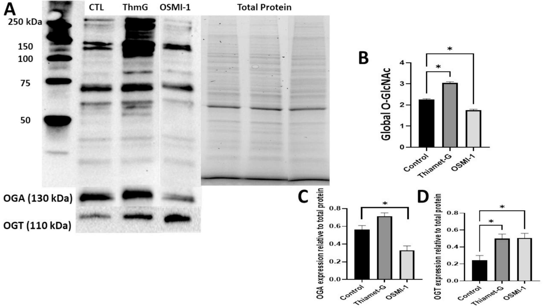 Fig. 1. Detection of O-GlcNAcylation in KGN cells following treatment with the small molecule inhibitors, Thiamet-G and OSMI-1 (Maucieri, Abigail M., and David H. Townson. 2024).
Cisplatin Promoted KGN Cell Apoptosis and Decreased the Expression of FTO and YAP1
Chemotherapy, especially the use of cisplatin, has been demonstrated to promote the apoptosis of granulosa cells. To investigate the mechanism by which cisplatin affects KGN cell apoptosis, flow cytometry analysis was used, and the results showed that cisplatin promoted KGN cell apoptosis in a dose-dependent manner (0-10 µM) (Fig. 2A-B). Furthermore, a series of key proteins related to KGN cell survival were measured. Western blotting was performed for FTO, BAX, Bcl-2, cleaved-caspase3, MST1, LATS1, and YAP1 and for the ratio of p-MST1/MST1, p-LATS1/LATS1, and p-YAP1/YAP1. As shown in the Fig. 2C-M, with increasing concentrations of cisplatin, the protein expression level of FTO decreased significantly, and this change was accompanied by increases in the expression levels of BAX and cleaved-caspase3 and a decrease in the expression levels of Bcl-2. In addition, the expression of the YAP1 protein decreased significantly after the cisplatin treatment, whereas the relative ratio of p-YAP1/YAP1 was significantly increased. The kinase proteins of the Hippo signaling pathway, such as MST1 and LATS1, gradually decreased. The relative ratios of p-MST1/MST1 and p-LATS1/LATS1 were significantly increased. Consistent with the western blot results, immunofluorescence demonstrated that in cisplatin-induced injured KGN cells, the expression level of YAP1, which is mainly localized in the nucleus of proliferative granulosa cells, was downregulated significantly (Fig. 2N - O). These results indicated that FTO was downregulated or inhibited may in a Hippo/YAP signaling pathway-dependent manner after cisplatin treatment.
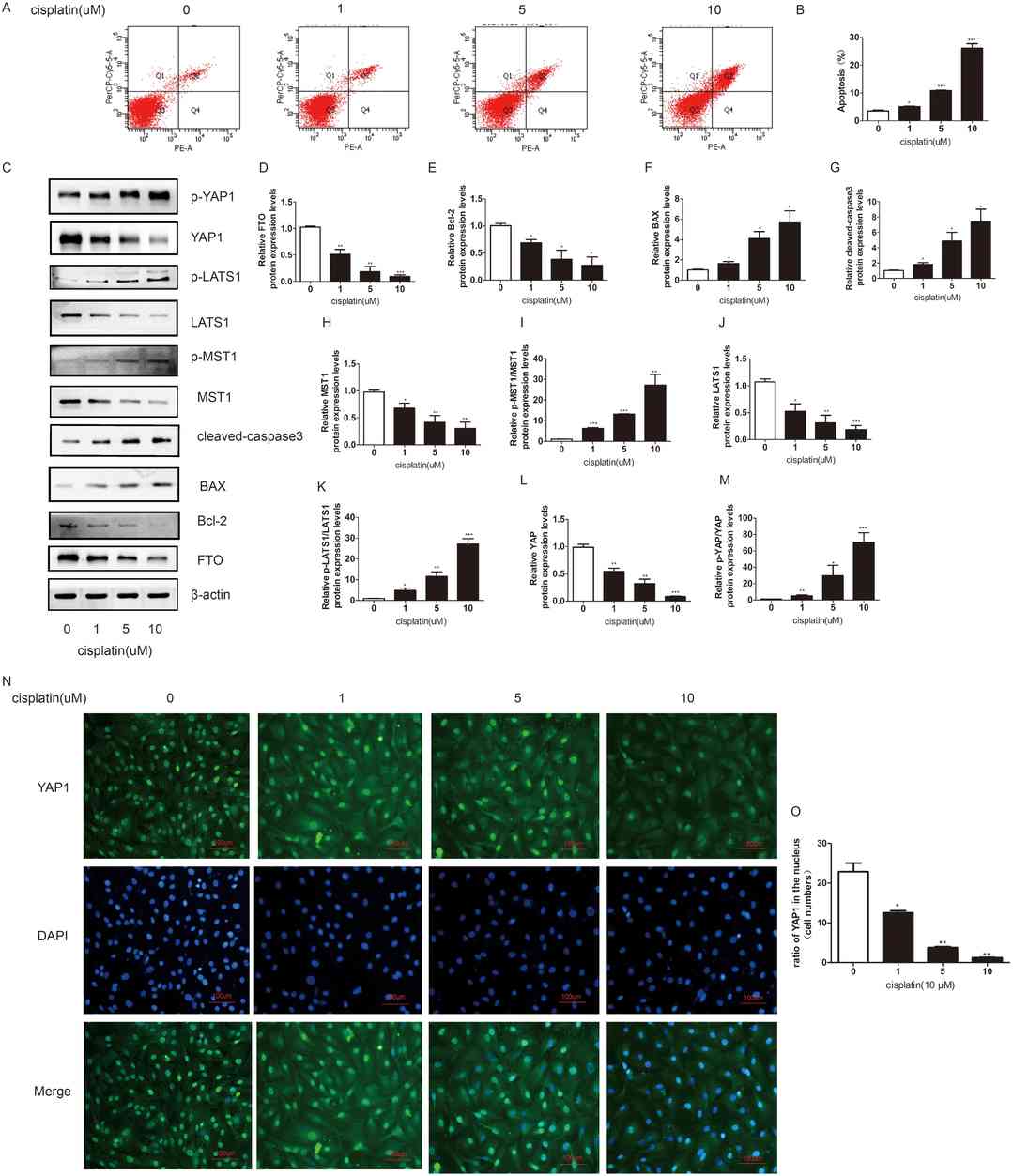 Fig. 2. Cisplatin promoted the KGN cell apoptosis, decreased the expression of FTO, and activated the Hippo/YAP signaling pathway (Wang, Rongli, et al. 2024).
The most common form of ovarian cancer begins in epithelial cells, which are the cells that line the surfaces and cavities of the body. These cancers can arise in the epithelial cells on the surface of the ovary.
Ask a Question
Average Rating: 5.0 | 1 Scientist has reviewed this product
Detailed protocols and guidelines
We appreciate the detailed protocols and guidelines provided alongside the tumor cell products.
18 Mar 2023
Ease of use
After sales services
Value for money
Write your own review
- You May Also Need
Description: Species: human female 60 years old;
Tissue: ovary;
Tumor: adenocarcinoma, low differentiated;
G3 FIGO stage IV (pleural metastasis);
Derived from: ascitic fluid

Description: Established from the solid omental metastasis of a mucinous papillary adenocarcinoma of the ovary of a 36-year-old woman in 1979; initially diploid, later tetraploid; growth in nude mice

Description: Established from the ascites fluid stemming from the colon metastasis of a 46-year-old white woman with ovarian adenocarcinoma in 1986

Description: Established from the ascitic fluid of a 56-year-old Caucasian woman with dedifferentiated serous cystadenocarcinoma of the ovary in 1979

Description: Established post-hysterectomy from the tumor tissue of a 65-year-old Japanese woman with malignant ovarian carcinoma (stage: FIGO IIIc; histology: poorly differentiated serous papillary adenocarcinoma) in 1989

Description: The cells are distributed for research purposes only. The culture condition is 37℃,95% air, carbon dioxide (CO2), complete growth medium supplemented with 5% (v/v) DMSO
