Featured Products
Our Promise to You
Guaranteed product quality, expert customer support

ONLINE INQUIRY
ELF-153
Cat.No.: CSC-C0674
Species: Human
Source: acute myeloid leukemia
Morphology: round cells growing singly in suspension; occasional semi adherent cells present; a few giant cells present
Culture Properties: suspension
- Specification
- Background
- Scientific Data
- Q & A
- Customer Review
Immunology: CD3 -, CD4 +, CD13 +, CD14 -, CD15 -, CD19 -, CD33 +, CD34 +, HLA-DR +
Viruses: PCR: EBV -, HBV -, HCV -, HIV -, HTLV-I/-II -
The ELF-153 cell line represents a human acute myeloid leukemia (AML) cell line created in 1988 from the bone marrow samples of a 41-year-old male. The patient began with acute myelofibrosis before progressing to develop refractory relapsed acute myeloid leukemia (AML-M7). This cell line shows normal megakaryocytic characteristics through its presence of both undifferentiated precursor cells and mature megakaryocytes. This cell line requires multiple growth factors for survival mainly IL-3, GM-CSF and GM-CSF-like cytokines. 5-azacytidine and phorbol ester treatment enables these cells to proliferate indefinitely while maintaining their differentiation characteristics.
ELF-153 has various biological functions. The supernatant from ELF-153 cells has been confirmed to enhance NRK-49F fibroblast growth and bovine endothelial cell development while prompting HEL cells to release interleukin-6 (IL-6). Subcutaneous injection of ELF-153 cells into nude mice produces hard tumors that show extensive blast infiltration and significant reticular fibrosis upon histological examination. Additionally, the ELF-153 cell line divides into three different cell populations (CD34+/CD61-, CD34+/CD61+, CD34-/CD61+) through flow cytometry which enables researchers to study megakaryocyte differentiation through their unique proliferative properties and endoplasmic reticulum characteristics.
ERG+85High Cells Exhibit Regenerative Properties
Acute leukemia is a fast-progressing blood cancer with poor survival rates, mainly due to poorly characterized leukemia stem cells (LSCs) that resist treatments and cause relapse. Aqaqe et al. employ an ERG+85 enhancer-based fluorescent reporter system to investigate and characterize subpopulations of leukemia cells with stem cell-like properties.
To assess the stability of cellular states marked by ERG+85, they analyzed tagBFP dynamics in sorted tagBFPHigh and tagBFPNeg Jurkat and ELF153 cells. FACS revealed tagBFPHigh cells could regenerate populations of tagBFPHigh, tagBFPIntermediate, and tagBFPNeg cells. In contrast, tagBFPNeg cells failed to produce tagBFPHigh cells even after 30 days (Fig. 1). A similar distribution was observed when assessing ERG+85 activity in progeny from individual tagBFPHigh or tagBFPNeg cells in colony assays. ERG protein analysis showed tagBFPHigh Jurkat cells had 2- to 3-fold higher ERG protein levels than tagBFPNeg cells (Fig. 1). Notably, both cell types had similar viral integration numbers, dismissing biases or stochastic expression variations of tagBFP. The presence of varying tagBFP levels in tagBFPHigh progeny suggests a gradual regulation mechanism rather than an on/off switch for ERG+85 dynamics. To compare the regenerative potential of tagBFPHigh and tagBFPNeg cells, they conducted a methycellulose colony replating assay. TagBFPHigh ELF-153 cells formed more colonies than tagBFPNeg cells across all platings, with tagBFPNeg cells failing to form colonies by the third plating, indicating lower self-renewal potential (Fig. 2A). They assessed AML blasts' regenerative capacity using stroma-supported cultures and limiting dilution analysis (LDA). TagBFPHigh cells showed 1.7- to 23-fold higher culture initiation than tagBFPNeg cells and demonstrated significant proliferation, unlike tagBFPNeg cells (Fig. 2B-D). In primary AML cells, tagBFPHigh cells could regenerate cells with varying ERG+85 activity, while tagBFPNeg could not produce tagBFPHigh cells (Fig. 2B-D). These findings are consistent with those in reporter-expressing leukemia cell lines.
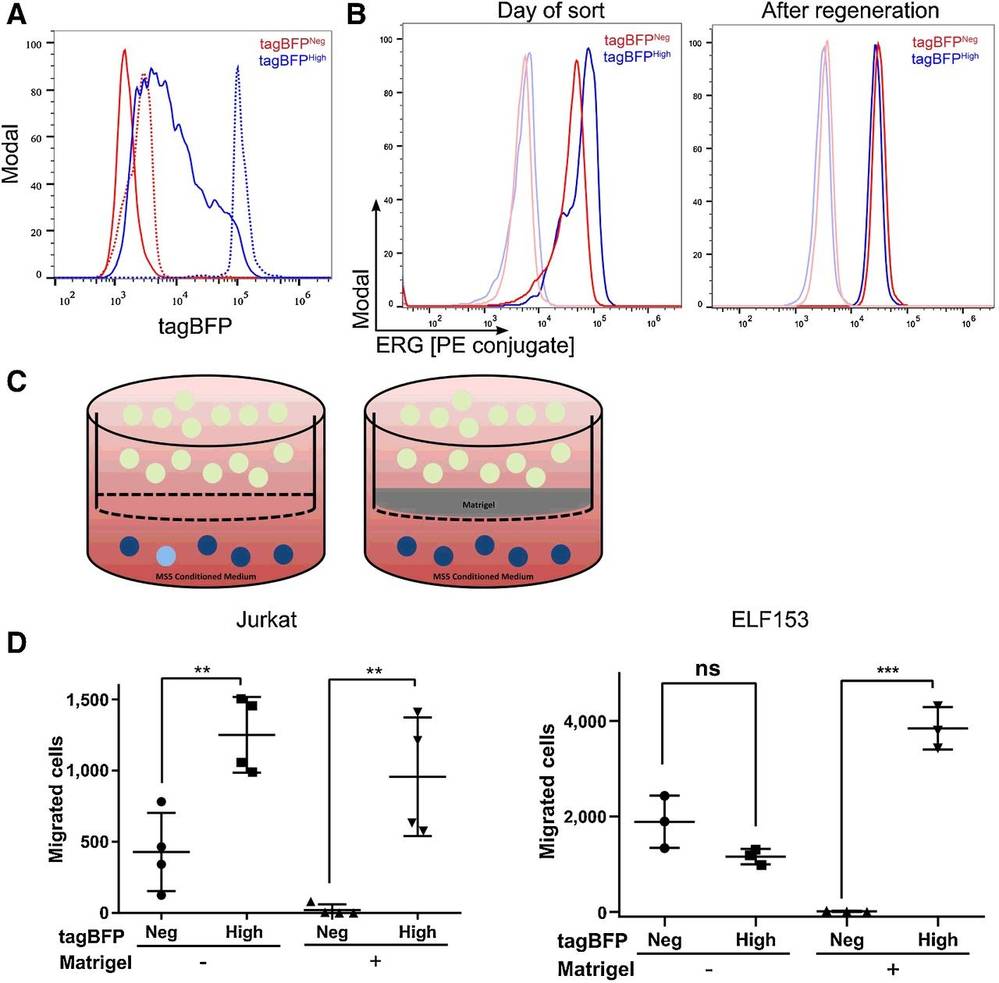 Fig. 1. Regeneration and migration/invasion potential of tagBFPHigh and tagBFPNeg fractions (Aqaqe N, Yassin M, et al., 2019).
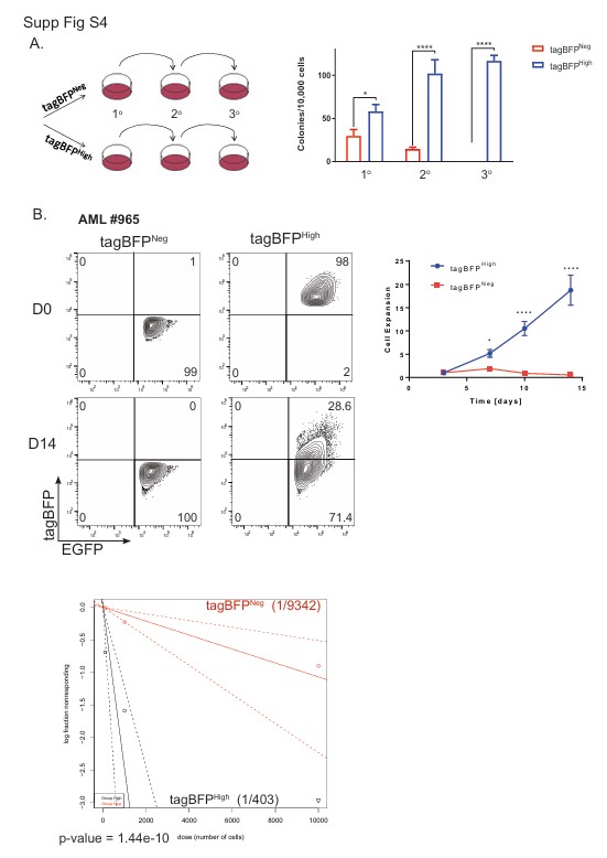 Fig. 2. ERG+85High cells exhibit regenerative properties (Aqaqe N, Yassin M, et al., 2019).
Aza-Caused Demethylation in Methylated CpG Sites of ELF-153 and MOLM-13 Cell Lines
Hypomethylating agents like 5-Azacytidine (Aza) are used to treat myeloid malignancies, improving survival in MDS, CMML, and AML patients. The exact molecular mechanisms behind the response to these agents remain unclear, especially since traditional treatment aims to eradicate malignant clones, yet Aza seems to stabilize clonal architecture instead.
Using next-generation sequencing (NGS), 30 myeloid leukemia-associated genes were analyzed in 15 MDS/CMML patients with excellent response to Aza. Gawliza et al. identified four main gene targets (LINE-1, EZH2, NOTCH1, RUNX1 and WT1) of Aza by methylation analysis. Then, in vitro evaluation of Aza response was conducted on ELF-153 and MOLM-13 cell lines. The 72-hour IC50 for Aza was 5.8 µmol/l for ELF-153 and 2.5 µmol/l for MOLM-13. They analyzed LINE-1, a global methylation marker, and potential target genes (LINE-1, NOTCH1, RUNX1 and WT1). After 72 hours of incubation, significant demethylation of LINE-1 and all genes was observed compared to untreated controls (0 h) (Fig. 3). Genes with higher baseline methylation (e.g., EZH2) showed greater absolute methylation reduction than those with lower baseline methylation (e.g., RUNX1). EZH2 exhibited the most significant reduction: 46.1% in ELF-153 and 37.5% in MOLM-13.
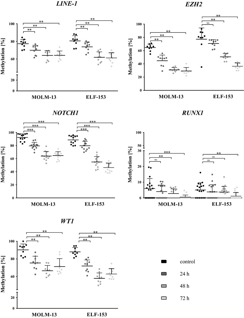 Fig. 3. Decrease in CpG methylation levels of LINE-1, EZH2, NOTCH1, RUNX1 and WT1 in MOLM-13 and ELF-153 incubated with Aza for 72 h (Gawliza A L, Speith J, et al., 2019).
Relatively inexpensive, fast turnaround time, highly sensitive, and high abundance detection range。
Ask a Question
Average Rating: 4.0 | 1 Scientist has reviewed this product
High quality
The quality of the tumor cell products from Creative Bioarray is backed by rigorous quality control measures, including cell viability and purity testing.
29 June 2022
Ease of use
After sales services
Value for money
Write your own review
- You May Also Need


Description: Species: human female; Tissue: lymphocyte, B; Pathology: paroxysmal nocturnal hemoglobinuria; Transformed by: Epstein-Barr
Description: Species: human female; Tissue: lymphocyte, B; Pathology: paroxysmal nocturnal hemoglobinuria; Transformed by: Epstein-Barr
Description: Species: human female; Tissue: lymphocyte, B; Pathology: paroxysmal nocturnal hemoglobinuria; Transformed by: Epstein-Barr

Description: Species: human male 66 years old;
Tissue: lymph node, metastasis;
Tumor: melanoma
Description: Species: human, Black male 11 year old;
Tumor: lymphoma, Burkitt;
Derived from: Raji cell line
