- You are here: Home
- Resources
- Explore & Learn
- ISH/FISH
- Whole Chromosome Painting Probes for FISH
Support
-
Cell Services
- Cell Line Authentication
- Cell Surface Marker Validation Service
-
Cell Line Testing and Assays
- Toxicology Assay
- Drug-Resistant Cell Models
- Cell Viability Assays
- Cell Proliferation Assays
- Cell Migration Assays
- Soft Agar Colony Formation Assay Service
- SRB Assay
- Cell Apoptosis Assays
- Cell Cycle Assays
- Cell Angiogenesis Assays
- DNA/RNA Extraction
- Custom Cell & Tissue Lysate Service
- Cellular Phosphorylation Assays
- Stability Testing
- Sterility Testing
- Endotoxin Detection and Removal
- Phagocytosis Assays
- Cell-Based Screening and Profiling Services
- 3D-Based Services
- Custom Cell Services
- Cell-based LNP Evaluation
-
Stem Cell Research
- iPSC Generation
- iPSC Characterization
-
iPSC Differentiation
- Neural Stem Cells Differentiation Service from iPSC
- Astrocyte Differentiation Service from iPSC
- Retinal Pigment Epithelium (RPE) Differentiation Service from iPSC
- Cardiomyocyte Differentiation Service from iPSC
- T Cell, NK Cell Differentiation Service from iPSC
- Hepatocyte Differentiation Service from iPSC
- Beta Cell Differentiation Service from iPSC
- Brain Organoid Differentiation Service from iPSC
- Cardiac Organoid Differentiation Service from iPSC
- Kidney Organoid Differentiation Service from iPSC
- GABAnergic Neuron Differentiation Service from iPSC
- Undifferentiated iPSC Detection
- iPSC Gene Editing
- iPSC Expanding Service
- MSC Services
- Stem Cell Assay Development and Screening
- Cell Immortalization
-
ISH/FISH Services
- In Situ Hybridization (ISH) & RNAscope Service
- Fluorescent In Situ Hybridization
- FISH Probe Design, Synthesis and Testing Service
-
FISH Applications
- Multicolor FISH (M-FISH) Analysis
- Chromosome Analysis of ES and iPS Cells
- RNA FISH in Plant Service
- Mouse Model and PDX Analysis (FISH)
- Cell Transplantation Analysis (FISH)
- In Situ Detection of CAR-T Cells & Oncolytic Viruses
- CAR-T/CAR-NK Target Assessment Service (ISH)
- ImmunoFISH Analysis (FISH+IHC)
- Splice Variant Analysis (FISH)
- Telomere Length Analysis (Q-FISH)
- Telomere Length Analysis (qPCR assay)
- FISH Analysis of Microorganisms
- Neoplasms FISH Analysis
- CARD-FISH for Environmental Microorganisms (FISH)
- FISH Quality Control Services
- QuantiGene Plex Assay
- Circulating Tumor Cell (CTC) FISH
- mtRNA Analysis (FISH)
- In Situ Detection of Chemokines/Cytokines
- In Situ Detection of Virus
- Transgene Mapping (FISH)
- Transgene Mapping (Locus Amplification & Sequencing)
- Stable Cell Line Genetic Stability Testing
- Genetic Stability Testing (Locus Amplification & Sequencing + ddPCR)
- Clonality Analysis Service (FISH)
- Karyotyping (G-banded) Service
- Animal Chromosome Analysis (G-banded) Service
- I-FISH Service
- AAV Biodistribution Analysis (RNA ISH)
- Molecular Karyotyping (aCGH)
- Droplet Digital PCR (ddPCR) Service
- Digital ISH Image Quantification and Statistical Analysis
- SCE (Sister Chromatid Exchange) Analysis
- Biosample Services
- Histology Services
- Exosome Research Services
- In Vitro DMPK Services
-
In Vivo DMPK Services
- Pharmacokinetic and Toxicokinetic
- PK/PD Biomarker Analysis
- Bioavailability and Bioequivalence
- Bioanalytical Package
- Metabolite Profiling and Identification
- In Vivo Toxicity Study
- Mass Balance, Excretion and Expired Air Collection
- Administration Routes and Biofluid Sampling
- Quantitative Tissue Distribution
- Target Tissue Exposure
- In Vivo Blood-Brain-Barrier Assay
- Drug Toxicity Services
Whole Chromosome Painting Probes for FISH
Fluorescence in situ hybridization (FISH) is a vital molecular cytogenetic technique in genetic research and clinical diagnosis. It allows the visualization of specific DNA sequences within chromosomes, aiding in detecting chromosomal abnormalities and genetic disorders. Whole chromosome painting probes play a crucial role in FISH by enabling the simultaneous visualization of entire chromosomes, providing valuable insights into the structural organization and behavior of chromosomes.

What are Whole Chromosome Painting Probes?
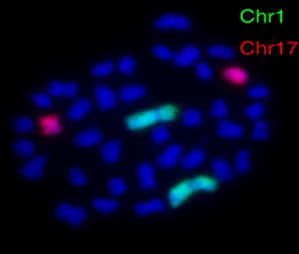
Whole chromosome painting probes are molecular tools designed to target and hybridize to the entire length of a specific chromosome. These probes are labeled with fluorescent dyes, allowing for the visualization and identification of the targeted chromosome within a cell nucleus. The development of whole chromosome painting probes involves various methods, including chromosome microdissection, degenerate oligonucleotide-primed polymerase chain reaction (DOP-PCR), and fluorescence-activated chromosome sorting (FACS).
Advantages of Whole Chromosome Painting Probes for FISH
- Comprehensive chromosomal analysis. Unlike traditional banding techniques, whole chromosome painting probes offer a comprehensive view of entire chromosomes, enabling the simultaneous visualization of chromosomal aberrations and structural rearrangements. This comprehensive analysis enhances the efficiency and accuracy of cytogenetic investigations, particularly in complex karyotypes and mosaicism.
- High sensitivity and specificity. Whole chromosome painting probes demonstrate high sensitivity and specificity in detecting chromosomal abnormalities, making them invaluable in both research and clinical settings. The precise targeting of entire chromosomes minimizes the risk of false-positive or false-negative results, ensuring the reliability of FISH-based analyses.
- Enhanced throughput and efficiency. Using whole chromosome painting probes streamlines the FISH workflow by simplifying the process of identifying and analyzing multiple chromosomal regions in a single experiment. This improved throughput and efficiency are particularly advantageous in high-volume cytogenetics laboratories, where rapid and accurate results are essential for patient care and clinical decision-making.
Whole Chromosome Painting Probes in Cytogenetics
Whole chromosome painting probes are used to detect chromosomal abnormalities such as aneuploidy (the presence of an abnormal number of chromosomes), translocations (rearrangements of parts between nonhomologous chromosomes), and deletions.
Whole Chromosome Painting Probes in Cancer Research
In the field of oncology, whole chromosome painting probes have emerged as indispensable tools for investigating chromosomal aberrations in cancer cells. By using these probes in FISH analysis, researchers can identify specific chromosomal changes associated with various types of cancer, aiding in tumor classification, prognostic assessment, and treatment decision-making.
Whole Chromosome Painting Probes in Genetic Disorders Identification
Whole chromosome painting probes also find extensive applications in the diagnosis of genetic disorders such as Down syndrome, Turner syndrome, and Klinefelter syndrome. Through FISH analysis with chromosome-specific painting probes, researchers can accurately detect aneuploidies and other chromosomal abnormalities.
Creative Bioarray Relevant Recommendations
Creative Bioarray has a full range of DNA probe libraries that use fluorescently labeled specific chromosomes (whole chromosome, specific chromosome arm, or chromosome fragments).
| Cat. No. | Product Name |
| FWCP-01 | WCP 1 FISH Probe |
| FWCP-02 | WCP 2 FISH Probe |
| FWCP-03 | WCP 3 FISH Probe |
| FWCP-04 | WCP 4 FISH Probe |
| FWCP-05 | WCP 5 FISH Probe |
| FWCP-06 | WCP 6 FISH Probe |
| FWCP-07 | WCP 7 FISH Probe |
| FWCP-08 | WCP 8 FISH Probe |
| FWCP-09 | WCP 9 FISH Probe |
| FWCP-10 | WCP 10 FISH Probe |
| FWCP-23 | WCP X FISH Probe |
| FWCP-24 | WCP Y FISH Probe |
View our full range of whole chromosome painting probes and find what you need!
For research use only. Not for any other purpose.

