What Types of Multicolor FISH Probe Sets Are Available?
The field of cytogenetics has long been interested in studying the chromosomes, which carry the genetic information of an organism. Traditionally, techniques like G-banding and karyotyping have been used to observe and analyze the chromosomes. However, these methods have limitations in their ability to detect complex chromosomal rearrangements and provide detailed information about specific chromosomal regions.
Multicolor FISH (mFISH) overcomes these limitations by employing a combination of fluorescently labeled probes. Each probe in the mFISH probe set is designed to target a specific DNA sequence within the genome. By using different fluorophores, each probe can emit a unique color when visualized under a fluorescence microscope. This allows researchers to simultaneously visualize multiple chromosomal regions within a single experiment.
What Are mFISH Probe Sets?
mFISH also known as spectral karyotyping (SKY), is a technique that utilizes a combination of fluorescently labeled probes to visualize specific chromosomal regions. Each fluorescent probe is designed to bind to a unique DNA sequence within the genome, and the resulting pattern of fluorescence allows for the identification and analysis of individual chromosomes or chromosomal regions.
mFISH probe sets consist of a collection of fluorescently labeled probes that target specific chromosomal regions. These probes are designed to emit distinct colors when visualized under a fluorescence microscope. By using different fluorophores, each probe set can be assigned a unique color, enabling the simultaneous visualization of multiple chromosomal regions within a single experiment.
Available Types of mFISH Probe Sets
- Whole chromosome painting mFISH probe sets. This technique separates cells based on their density using a density gradient medium. It allows for the separation of PBMCs or dissociated tissue cells from other components, resulting in a fraction enriched in NK cells. The advantage of whole chromosome painting mFISH probe sets is their ability to detect complex chromosomal rearrangements, such as translocations and inversions, which may involve multiple chromosomes. By visualizing the entire chromosome, researchers can accurately assess the structural integrity of the genome and identify any abnormalities.
- mFISH banding probe sets. They are designed to generate a banded pattern along the length of each chromosome. These probe sets consist of multiple probes that bind to specific chromosomal bands, which are regions of the chromosome that have characteristic staining patterns when treated with specific dyes. The use of mFISH banding probe sets allows for the identification of chromosomal bands, which are useful for cytogenetic analysis and karyotyping. The patterns of bands can provide valuable information about the structural organization of the chromosomes and aid in the identification of specific chromosomal aberrations.
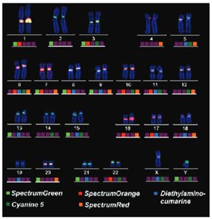
- Centromere and/or locus-specific mFISH probe sets. They are designed to target specific regions of the chromosomes, such as centromeres or specific gene loci. These probe sets consist of probes that bind to specific DNA sequences within the targeted regions, enabling the visualization of these regions with distinct colors. The use of centromere and/or locus-specific mFISH probe sets allows for the precise identification and analysis of specific chromosomal regions of interest. This is particularly useful in studies focused on centromere abnormalities, gene amplifications, or specific genomic rearrangements.
 Fig.1 Centromere-specific multicolor FISH (cenM-FISH) on a normal male metaphase. (Nietzel A, et al., 2001)
Emerging Technologies in mFISH Probe Sets
- Super-resolution FISH. Super-resolution FISH techniques employ various methods to surpass the diffraction limit, such as stimulated emission depletion (STED) microscopy, structured illumination microscopy (SIM), and single-molecule localization microscopy (SMLM). These techniques utilize advanced optics, fluorophores, and computational algorithms to achieve enhanced resolution. Super-resolution FISH enables imaging at a resolution beyond the diffraction limit of conventional microscopy, typically reaching down to tens of nanometers. This enhanced resolution allows for the visualization of intricate molecular structures and spatial relationships within the cell, providing insights into cellular organization and function.
- Multiplexed FISH. Multiplexed FISH techniques employ various strategies to detect multiple targets within the same sample. These strategies include the use of spectrally distinct fluorophores, DNA barcode-based approaches, sequential hybridization and imaging, and multiplexed probe design using combinatorial labeling schemes. Multiplexed FISH allows the detection and localization of multiple genes, RNAs, or proteins within the same sample, providing insights into their coordinated expression, interactions, and spatial relationships. It enables the investigation of complex biological phenomena, such as gene regulatory networks, cell signaling pathways, and cellular heterogeneity.
Applications of mFISH Probe Sets
- Chromosomal abnormality detection. mFISH probe sets are widely used for the detection and characterization of chromosomal abnormalities associated with genetic disorders and cancer. By visualizing the entire genome in a single experiment, mFISH can identify numerical and structural chromosomal aberrations, such as aneuploidy, translocations, deletions, and duplications.
- Genomic mapping and comparative genomics. mFISH probe sets are instrumental in genomic mapping and comparative genomics studies. By providing a comprehensive view of the genome, mFISH allows researchers to compare chromosomal organization and structural variations between different species or individuals. This information aids in understanding the evolutionary relationships between species, detecting genomic rearrangements, and identifying conserved or lineage-specific chromosomal regions.
- Research in developmental biology and cell biology. mFISH probe sets have significant applications in developmental biology and cell biology research. They allow researchers to investigate the spatial and temporal organization of chromosomes during various stages of development or in different cell types. By visualizing chromosomal dynamics, researchers can gain insights into chromosomal behavior, nuclear organization, and the regulation of gene expression.
Creative Bioarray Relevant Recommendations
| Product/Service Types | Description |
| Multicolor FISH (M-FISH) Analysis | We provide multicolor FISH (M-FISH) assays for a precise assessment of complex chromosomal rearrangements. |
| Fluorescent In Situ Hybridization (FISH) Service | Creative Bioarray offers a full line of FISH services, from standardized testing of validated assays to custom development of new assays. |
| FISH Probe Design, Synthesis and Testing Service | Creative Bioarray frequently receives requests for custom synthesized probes, for novel, rare, and specialized applications. |
| ISH/FISH Probes | Creative Bioarray provides the most comprehensive list of FISH probes for the rapid identification of a wide range of chromosomal aberrations across the genome. |
References
- Nietzel A, et al. (2011). "A new multicolor-FISH approach for the characterization of marker chromosomes: Centromere-specific multicolor-FISH (cenM-FISH)." Hum Genet. 108, 199-204.
- Liehr T, et al. (2004). "Multicolor FISH probe sets and their applications." Histology and histopathology, 191, 229-37.