How to Use FISH in Hematologic Neoplasms?
Fluorescence in situ hybridization (FISH) is a molecular cytogenetic technique developed in the 1980s to detect specific DNA sequences on chromosomes. FISH uses fluorescent probes attached to the DNA target to detect chromosomal variations without harming the integrity of the cell. For use in medicine, FISH has been deployed extensively to find mutations in genes, chromosome changes and disease markers, directly emerging from the intersection of cytogenetics and molecular biology. One of the advantages of FISH is that chromosomal and genetic aberrations are found in real time, in the cell nucleus, making it fast and highly sensitive. This property has been crucial in diagnosing and characterizing hematological malignancies where FISH has the greatest potential.
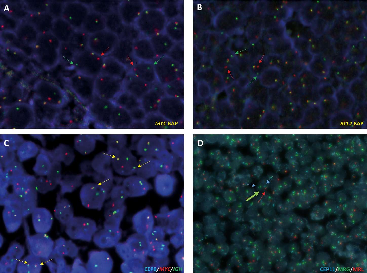
 Fig. 1. FISH analysis of 14q32 (IGH)-associated translocations and 11q aberration in B-cell lymphomas on formalin-fixed paraffin-embedded (FFPE) tissues (Salaverria I, Siebert R, et al., 2024).
FISH Applications in Hematologic Neoplasms Diagnosis and Treatment
- Auxiliary diagnosis and disease classification: FISH is used to confirm the diagnosis of certain blood diseases by identifying specific genetic abnormalities.
- Guidance for drug selection: Results of FISH analysis can be used as a basis for individualized medication strategies. For targeted drug therapies, FISH helps clinicians make the correct selection based on identified gene rearrangements or amplifications, and determine therapeutic efficacy.
- Treatment monitoring and evaluation: During treatment, FISH monitors changes in copy number of marker genes, providing insight into the response to treatment and residual disease status. FISH is sensitive enough to recognise minimal residual disease in leukemia and provide insight into treatment efficacy and relapse potential.
- Implantation status monitoring: In allogeneic hematopoietic stem cell transplantation, FISH determines the outcome of a transplant by tracking sex chromosome changes and aiding in post-transplant treatment and relapse prediction.
Advantages of FISH Technique in Hematologic Neoplasms
- High sensitivity and specificity: FISH detects subtle deletions or translocations that remain undetected in traditional karyotype analysis.
- Interphase cell detection: Unlike conventional cytogenetic testing that requires metaphase cells, FISH detects chromosomal anomalies in interphase cell nuclei without the need for cell culturing, streamlining result interpretation and acceleration.
- Wide specimen applicability: FISH is compatible with a diverse range of sample types, including peripheral blood, bone marrow smears, and paraffin-embedded tissue. This versatility grants FISH unique benefits in detecting rare mutations in biopsy samples, where sample processing poses a challenge.
- Rapid and quantifiable results: While typical karyotype analysis can take several days, FISH delivers results within 24 hours, significantly expediting diagnoses for conditions like promyelocytic leukemia.
- Simultaneous multi-genomic target analysis: A single FISH experiment can analyze multiple genomic targets, providing comprehensive diagnostic insight in complex cases.
- Detection of specific cryptic chromosomal abnormalities.
When to Apply FISH Technology?
- When clinical signs indicate specific chromosomal aberrations, such as the (t(8;14) in Burkitt lymphoma, the MYC/IGH probe can be used to determine them.
- For risk assessment and disease control, specifically in acute and chronic lymphoblastic leukemia and multiple myeloma, FISH is used to assess genetic abnormalities and treatment.
- When standard cytogenetic testing fails to detect defects. For example, the t(12;21) translocation in acute lymphoblastic leukemia.
- As a marker for minimal residual disease, FISH assists in evaluate treatment efficacy and early recurrence in the context of chemotherapy and hematopoietic stem cell transplantation.
- Paraffin-embedded tissue slices identify chromosomal translocations in lymphoma.
- Follow-up of cross-gender patients receiving bone marrow transplants involves monitoring chimerism status to determine the transplant's efficacy and guide future care.
- Rapid diagnosis for acute promyelocytic leukemia is achieved with FISH which quickly identifies the PML-RARA fusion gene, leading to rapid diagnosis within 2-3 hours.
Application Example
- Acute myeloid leukemia (AML): FISH is very useful for the genetic diagnosis and prognosis prediction of AML, i.e., detection of AML1/ETO, PML-RARa, MLL gene break rearrangements, etc.
- Chromosomal abnormality (CLL): The chromosomal abnormality in CML is t(9;22)(q34;q11.2), which creates the BCR/ABL fusion gene. This fusion gene can be identified using FISH, diagnosing CML.
- Multiple myeloma (MM): FISH has a special benefit when it comes to multiple myeloma because it is able to detect chromosomal abnormalities (1q21 amplification, p53 deletion, and 13q14 deletion) in the presence of low plasma cell number and complex chromosomal abnormalities.
- Lymphoma: FISH can identify gene mismatches in lymphoma, determining the nature of the disease and aiding in the assessment of the disease's nature and potential response to therapy.
- Myelodysplastic syndromes (MDS): For MDS, FISH replaces the failures of traditional karyotype identification and enhances the sensitivity and accuracy of early detection with cytogenetics.
Sensitivity, rapidity and broad flexibility made FISH an integral part of diagnosing and studying hematological malignancies. With new technologies advancing and app protocols evolving, FISH will be able to provide even more efficient and precise services for hematological tumor patients in the future.
| Products & Services | Description |
| Fluorescent In Situ Hbridization (FISH) Service | Creative Bioarray offers a range of different FISH services including metaphase and interphase FISH, fibre-FISH, RNA-FISH, M-FISH, 3D-FISH, flow-FISH, FISH on paraffin sections, and immune-FISH. |
| Neoplasms FISH Analysis | Creative Bioarray offers a wide menu of probes for fluorescent in situ hybridization (FISH) testing. FISH can help identify subtle or sub-microscopic structural rearrangements, variant chromosomes, and low-frequency abnormalities not readily detectable by class cytogenetics. |
Reference
- Salaverria I, Siebert R, et al. Appraisal of current technologies for the study of genetic alterations in hematologic malignancies with a focus on chromosome analysis and structural variants. Med Genet. 2024, 6;36(1),13-20.