How Important are Lipids in Exosome Composition and Biogenesis?
The lipids of the exosomes include cholesterol, SM, glycosphingolipids and phosphatidylserine. These lipids are not only the basis of exosome structure, but also involved in the formation and release of exosomes. Additionally, it regulates exosome biogenesis, release, targeting and cellular adhesion.
Even though lipidomics is somewhat less studied than genomics and exosome proteomics. Yet interest in exosome lipidomics has picked up dramatically in recent years. Since 2006, there has been a 15-fold increase in related studies. As lipidomics has grown, the role of lipids in exosome biology, and specifically in understanding the potential for exosomes as biomarkers and therapeutic targets, becomes increasingly evident.
Exosome Lipid Composition
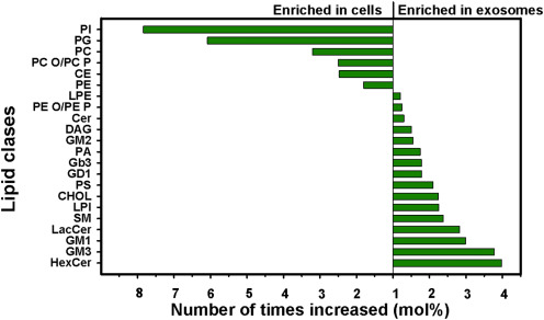
Similar to the common lipid composition of cell membranes, sphingolipids, phospholipids, ganglioside GM3, and cholesterol are also major lipid classes in exosomes. However, cholesterol, sphingomyelin, sphingolipid, and phosphatidylserine were 2-to 3-fold enriched from cells to exosomes. In contrast, exosomes generally contained less PC (mol% of total lipids) than their parent cells, and only small changes were reported for PE.
 Fig. 1. Changes in the relative amounts of lipid classes from intracellular to exosomal in PC-3 cells (Record M, Silvente-Poirot S, et al., 2018).
Furthermore, there are differences in different exosomes, varying in abundance based on cell type, physiological conditions, and exosome functions. For example, exosomes from different sources show distinct lipid enrichments, like high cholesterol in B-lymphocyte-derived exosomes, impacting exosome stability and interactions. In addition, the membrane of exosomes of PC3 cell origin is a highly ordered structure rich in sphingolipids, which confers the stability that exosomes exhibit in the extracellular environment. Table 1 lists some relevant studies on the differential distribution of lipids in exosomes from different sources.
Table. 1. Lipid composition of exosomes from different sources.
| Exosome source | Enriched lipids | Abbreviations |
| B16-F10 | CerG2, CL, LPG, Cer, TG. | Cer, ceramide; CL, cardiolipin; Cer, ceramide; CerG1-3, glucosylceramides; CerP, Ceramide phosphates; DAG, diacylglycerol; FFA, free fatty acids; LPC, lysophosphatidylcholine; LPG, lysophosphatidylglycerol; M(IP)2C, Mannosyl-di-PI-ceramides; MG, monoglyceride; PG, phosphatidylglycerol; PI, phosphatidylinositol; SHexCer, Sulfatides hexosyl ceramide; SHexSph, Sulfatides hexosyl sphingoid bases; TG, triglyceride. |
| MDA-MB-231 | MG, CerG2; Cer, LPG; LPC; PG; TG; CL. | |
| AsPC-1 | DAG, TG, LPC, Cer, CerG1, LPG, MG, PG. | |
| Human Urine | CerP, HexSph, LacCer, M(IP)2C, SHexCer, SHexSph. | |
| U87 | Glycolipids, FFA, CL. | |
| Mesenchymal Stem Cells | Glycolipids, FFA, CL. |
The Role of the Lipids in Exosome Biogenesis
Exosome biogenesis can be understood as a three-step process: the first step is the biogenesis of MVBs, followed by the transport of MVBs to the plasma membrane, and finally the release of MVBs from luminal vesicles by fusion to the plasma membrane.
Cholesterol: Cholesterol plays a critical role in exosome assembly, because it assists the ESCRT machinery to create MVBs and stuff cargo into exosomes. The presence of cholesterol within the exosome membrane allows for the arrangement of microdomains in the membrane that is needed for the assembly of exosomes.
Sphingolipids: Ceramides are derived from sphingolipids, which have a unique conical structure which promotes negative membrane curvature, initiating escrt-independent pathways that induce spontaneous membrane invagination. It has been shown in vitro that ceramides can independently induce membrane outgrowth and vesicle formation.
Phospholipids: Phospholipids: Phosphatidic acid (PA), the simplest phospholipid, is similar in structure to ceramides and also has the capacity to produce spontaneous negative membrane curvature. It has been reported that PA interacts with certain proteins to promote the budding of intraluminal vesicles (ILVs). In addition, it may also combine with sphingomyelinase to increase ceramide production and enhance ILV budding via an escrt-independent pathway.
Table.2. Role of relevant lipids during exosome biogenesis (Donoso-Quezada J, Ayala-Mar S, et al., 2021).
| Lipid types | Process involved | Role in exosome biogenesis |
| Cholesterol | EVs formation, transport and release. |
|
| Ceramide | EVs formation |
|
| Diacylglycerol | EVs formation |
|
| Ether lipids | EVs release |
|
| Phosphatidic acid | EVs formation |
|
| Phosphatidylinositol 3-phosphate | EVs formation and cargo sorting. |
|
| Bis(monoacyl-glycero) phosphate | EVs formation and release. |
|
| Cardiolipin | EVs stabilization |
|
| Phosphatidylinositol-3,5-biphosphate | EVs release |
|
| Sphingosine 1-phosphate | Cargo sorting |
|
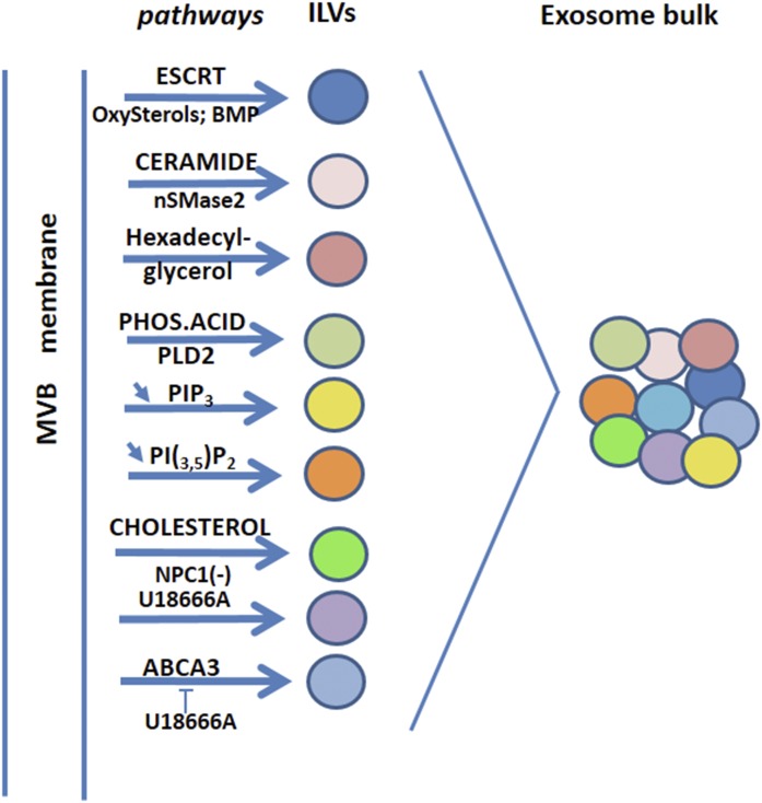
In addition, many lipid-related pathways are involved in exosome biogenesis. The mobilization of these pathways may depend on the type of parental cell, the nature of the initial stimulus, and the microenvironment, which makes it possible for the biogenesis pathways to combine to generate vesicles with different contents.
 Fig. 2. Lipid pathways involved in exosome biogenesis processes (Record M, Silvente-Poirot S, et al., 2018).
Exosomal lipids have received widespread interest for their potential medical applications over the past few years. Exosomal lipids are not only considered biomarkers for developing non-invasive diagnostic methods but are also seen as potential therapeutic targets for controlling disease progression and pathogenesis. These small vesicles help to keep cells in touch, and their lipid profile can control the growth and development of various pathologies. An emerging field, exosome lipidomics, is concerned with measuring the changes in lipids of exosomes under pathological conditions. This research helps us better grasp how lipids interact with cellular communication, and how they play a role in the driving forces behind disease pathogenesis.
| Products & Services | Description |
| Exosome Lipidomics Service | Creative Bioarray makes it easy to perform your exosome lipidomics research. Our high-throughput lipidomics analysis can provide lipid identification and quantification of your exosomes in detail. |
References
- Record M, Silvente-Poirot S, et al. Extracellular vesicles: lipids as key components of their biogenesis and functions. J Lipid Res. 2018 Aug;59(8):1316-1324.
- Donoso-Quezada J, Ayala-Mar S, et al. The role of lipids in exosome biology and intercellular communication: function, analytics and applications. Traffic. 2021.

