- You are here: Home
- Resources
- Explore & Learn
- Exosome
- Emerging Technologies and Methodologies for Exosome Research
Support
-
Cell Services
- Cell Line Authentication
- Cell Surface Marker Validation Service
-
Cell Line Testing and Assays
- Toxicology Assay
- Drug-Resistant Cell Models
- Cell Viability Assays
- Cell Proliferation Assays
- Cell Migration Assays
- Soft Agar Colony Formation Assay Service
- SRB Assay
- Cell Apoptosis Assays
- Cell Cycle Assays
- Cell Angiogenesis Assays
- DNA/RNA Extraction
- Custom Cell & Tissue Lysate Service
- Cellular Phosphorylation Assays
- Stability Testing
- Sterility Testing
- Endotoxin Detection and Removal
- Phagocytosis Assays
- Cell-Based Screening and Profiling Services
- 3D-Based Services
- Custom Cell Services
- Cell-based LNP Evaluation
-
Stem Cell Research
- iPSC Generation
- iPSC Characterization
-
iPSC Differentiation
- Neural Stem Cells Differentiation Service from iPSC
- Astrocyte Differentiation Service from iPSC
- Retinal Pigment Epithelium (RPE) Differentiation Service from iPSC
- Cardiomyocyte Differentiation Service from iPSC
- T Cell, NK Cell Differentiation Service from iPSC
- Hepatocyte Differentiation Service from iPSC
- Beta Cell Differentiation Service from iPSC
- Brain Organoid Differentiation Service from iPSC
- Cardiac Organoid Differentiation Service from iPSC
- Kidney Organoid Differentiation Service from iPSC
- GABAnergic Neuron Differentiation Service from iPSC
- Undifferentiated iPSC Detection
- iPSC Gene Editing
- iPSC Expanding Service
- MSC Services
- Stem Cell Assay Development and Screening
- Cell Immortalization
-
ISH/FISH Services
- In Situ Hybridization (ISH) & RNAscope Service
- Fluorescent In Situ Hybridization
- FISH Probe Design, Synthesis and Testing Service
-
FISH Applications
- Multicolor FISH (M-FISH) Analysis
- Chromosome Analysis of ES and iPS Cells
- RNA FISH in Plant Service
- Mouse Model and PDX Analysis (FISH)
- Cell Transplantation Analysis (FISH)
- In Situ Detection of CAR-T Cells & Oncolytic Viruses
- CAR-T/CAR-NK Target Assessment Service (ISH)
- ImmunoFISH Analysis (FISH+IHC)
- Splice Variant Analysis (FISH)
- Telomere Length Analysis (Q-FISH)
- Telomere Length Analysis (qPCR assay)
- FISH Analysis of Microorganisms
- Neoplasms FISH Analysis
- CARD-FISH for Environmental Microorganisms (FISH)
- FISH Quality Control Services
- QuantiGene Plex Assay
- Circulating Tumor Cell (CTC) FISH
- mtRNA Analysis (FISH)
- In Situ Detection of Chemokines/Cytokines
- In Situ Detection of Virus
- Transgene Mapping (FISH)
- Transgene Mapping (Locus Amplification & Sequencing)
- Stable Cell Line Genetic Stability Testing
- Genetic Stability Testing (Locus Amplification & Sequencing + ddPCR)
- Clonality Analysis Service (FISH)
- Karyotyping (G-banded) Service
- Animal Chromosome Analysis (G-banded) Service
- I-FISH Service
- AAV Biodistribution Analysis (RNA ISH)
- Molecular Karyotyping (aCGH)
- Droplet Digital PCR (ddPCR) Service
- Digital ISH Image Quantification and Statistical Analysis
- SCE (Sister Chromatid Exchange) Analysis
- Biosample Services
- Histology Services
- Exosome Research Services
- In Vitro DMPK Services
-
In Vivo DMPK Services
- Pharmacokinetic and Toxicokinetic
- PK/PD Biomarker Analysis
- Bioavailability and Bioequivalence
- Bioanalytical Package
- Metabolite Profiling and Identification
- In Vivo Toxicity Study
- Mass Balance, Excretion and Expired Air Collection
- Administration Routes and Biofluid Sampling
- Quantitative Tissue Distribution
- Target Tissue Exposure
- In Vivo Blood-Brain-Barrier Assay
- Drug Toxicity Services
Emerging Technologies and Methodologies for Exosome Research
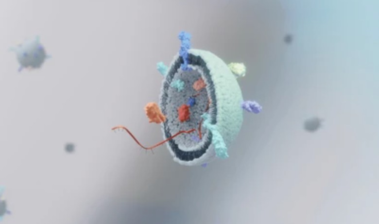
Exosomes, a subset of extracellular vesicles, have garnered significant attention in the field of biomedical research due to their diverse functions and potential applications in diagnostics, therapeutics, and regenerative medicine. As the demand for exosome research continues to grow, innovative technologies and methodologies are emerging to overcome existing challenges and expand our understanding of these fascinating nanosized vesicles.

High-Resolution Imaging Techniques
Advances in high-resolution imaging techniques have revolutionized the visualization and characterization of exosomes. These techniques enable researchers to study exosome morphology, cargo distribution, and cellular interactions at the nanoscale level.
- Electron microscopy (EM). Transmission electron microscopy (TEM) and scanning electron microscopy (SEM) allow for detailed visualization of exosome ultrastructure, providing insights into their size, shape, and membrane characteristics.
- Atomic force microscopy (AFM). AFM enables the imaging of exosomes in their native state, allowing for high-resolution topographical mapping of individual vesicles and their surface properties.
- Super-resolution microscopy. Techniques such as stimulated emission depletion (STED) microscopy, structured illumination microscopy (SIM), and single-molecule localization microscopy (SMLM) enhance spatial resolution beyond the diffraction limit, enabling the visualization of exosome subcellular localization and molecular interactions.
Omics Approaches
Omics technologies have revolutionized the comprehensive characterization of exosomal cargo, including nucleic acids, proteins, and lipids. These approaches provide valuable insights into the molecular composition and functional significance of exosomes.
- Exosome RNA sequencing (RNA-Seq). RNA-Seq enables the profiling of exosomal RNA species, including messenger RNA (mRNA), microRNA (miRNA), and long non-coding RNA (lncRNA). This technique facilitates the identification of disease-specific RNA signatures and regulatory networks.
- Proteomics. Mass spectrometry-based proteomics enables the identification and quantification of proteins within exosomes. This approach allows for the characterization of exosomal protein cargo, post-translational modifications, and protein-protein interactions.
- Lipidomics. Lipidomics approaches, such as mass spectrometry-based lipid profiling, enable the comprehensive analysis of lipid species present in exosomes. This technology provides insights into lipid composition, metabolism, and signaling pathways associated with exosomal functions.
Single-Exosome Analysis
Single-exosome analysis technologies have emerged as powerful tools for studying the heterogeneity and functional diversity of exosomes at the individual vesicle level. These methodologies offer the ability to analyze and manipulate single exosomes, providing detailed information on their cargo and functional properties.
- Single-exosome flow cytometry. Flow cytometry-based approaches allow for the analysis of individual exosomes based on size, surface markers, and cargo content. This technology enables high-throughput characterization and isolation of specific exosome subsets.
- Microfluidics-based analysis. Microfluidics platforms provide precise control over fluid flow and enable the isolation, manipulation, and analysis of single exosomes. These systems allow for real-time imaging, molecular profiling, and functional assays at the single-vesicle level.
Creative Bioarray Relevant Recommendations
Creative Bioarray provides a complete set of products and services for exosome research and resolves your issues quickly and efficiently. We also offer comprehensive support for customized services to meet your needs.
| Product/Service Types | Description |
| Exosome Standards | Creative Bioarray provides the best quality lyophilized exosome standards obtained from several biological samples, including hundreds of different cell lines, plasma, serum, saliva, and urine as well as other bio-fluids. |
| Exosome Antibodies | Creative Bioarray offers a list of monoclonal/polyclonal antibodies against the common and disease-specific exosomal proteins. These antibodies are suitable for common applications including WB, ELISA, FC, IHC, ICC, IF, IP, etc. |
| Exosome Identification | Creative Bioarray provides comprehensive support for your exosome identification by including the morphology assay, purity, and quantity assay, particle size distribution analysis, and exosome-specific markers expression. |
| Exosome Quantification | Creative Bioarray offers a range of options to meet most exosome quantitation demands. We provide reliable, and optimized tools at the most competitive price for quantitative analysis of exosomes in diverse biological samples such as plasma, urine, etc. |
| Exosome Analysis | Creative Bioarray provides diverse exosomal species analysis to help you understand your exosome compositions. |
For research use only. Not for any other purpose.

