Featured Products
Our Promise to You
Guaranteed product quality, expert customer support

ONLINE INQUIRY
Lu99
Cat.No.: CSC-C6353J
Species: Human
Morphology: epithelial-like
Culture Properties: Adherent cells
- Specification
- Background
- Scientific Data
- Q & A
- Customer Review
Store in liquid nitrogen.
The LU99 cell line is a human lung giant cell carcinoma cell line that serves as a crucial model for studying this rare and aggressive subtype of lung cancer. Giant cell carcinoma, characterized by the presence of large, atypical cells, is a variant of non-small cell lung cancer (NSCLC) that typically exhibits a poor prognosis. The LU99 cell line was established to provide researchers with a platform to investigate the unique biological behaviors and molecular mechanisms associated with giant cell carcinoma, which are often distinct from those of more common lung cancer types.
Derived from a patient with lung cancer, LU99 retains many of the morphological and genetic characteristics of the original tumor. This includes the expression of specific markers associated with giant cell carcinoma, which allows for the examination of tumor growth, differentiation, and the underlying signaling pathways that drive malignancy. The study of LU99 cells has implications for understanding how this aggressive cancer develops, as well as the factors that contribute to its resistance to conventional therapies.
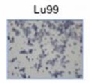 Fig. 1 Cell motility of LU99 cell line. (Rawangkan A, et al., 2022)
Dinactin Inhibited Tumor-Sphere Formation in Lu99 and A549 Cells
Lung cancer, especially NSCLC, is one of the most complex diseases, despite the existence of effective treatments such as chemotherapy and immunotherapy. Recently, dinactin has been revealed as a promising antitumor antibiotic via various mechanisms. However, the evidence relating to cell cycle progression regulation is constrained, and effects on cancer stemness have not been elucidated.
The inhibitory effects of dinactin on cancer stemness properties were investigated by tumor-sphere formation assay. Lu99 and A549 cells were cultured under a non-attached and serum-free CSC culture medium in the presence of 0.1 and 1 nM of dinactin for 14 days. The number of tumor-spheres greater than 100 µm was counted and kept for RNA extraction. As shown in Fig. 2a, b, non-treated Lu99 cells produced 195.5 ± 45.3 tumor-spheres (control), whereas treatment with 0.1 and 1 nM of dinactin inhibited tumor-sphere formation by 195.3 ± 39.3 and 9.33 ± 11.3 (95.2% inhibition), respectively. In A549 cells, similar results were obtained: treatment with 0.1 and 1 nM of dinactin inhibited tumor-sphere formation from 144.5 ± 69.5 to 109.7 ±15.9 (24.1% inhibition) and 41.0 ± 15.8 (71.6% inhibition), respectively. Furthermore, dinactin reduced the size of tumor spheres, as shown in Table 1. At a concentration of 1 nM of dinactin, the average size of tumor spheres was reduced from 175.54 ± 55.78 µm to 128.82 ± 20.07 µm (26.6% inhibition) in Lu99 cells and from 158.72 ± 47.40 µm to 134.87 ± 25.71 µm (15.0% inhibition) in A549 cells. These are new findings that highlight the very low concentration of dinactin (1 nM) capable of inhibiting CSC in NSCLC cells.
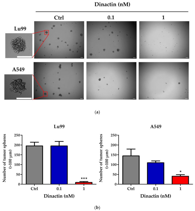 Fig. 2 Dinactin inhibited tumor-sphere formation in Lu99 and A549 cells. (Rawangkan A, et al., 2022)
Table 1 Dinactin reduced the size of tumor spheres in Lu99 and A549 cells. (Rawangkan A, et al., 2022)
| Dinactin (nM) | Average Size of Tumor Spheres (µm) | |
| Lu99 | A549 | |
| Control 0.1 1 | 175.54 ± 55.78 181.04 ± 46.43 128.82 ± 20.07 *** | 158.72 ± 47.40 159.28 ± 43.98 134.87 ± 25.71 ** |
Lu99 Cell Culture Supernatant Induces HDGF- and VEGF-Independent Tube Formation
Most NSCLC cells overexpress vascular endothelial growth factor-A (VEGF-A) which plays a pivotal role in tumor angiogenesis. Anti-angiogenic therapies including VEGF-A neutralization have significantly improved the response rates, progression-free survival, and overall survival of patients with NSCLC. To determine whether HDGF is involved in angiogenesis induced by the other NSCLC cell lines, a novel culture method was established; NSCLC cells were embedded in collagen gel and cultured three-dimensionally in a tube-induction medium containing FBS (Fig. 3A). The 3D culture supernatants derived from Lu99 cells incubated at 0-5×10⁶ cells/ml for 24 h significantly induced tube formation in a cell density-dependent manner, whereas no significant differences were noted in tube formation when using different cell densities (2-5×10⁶ cells/ml; Fig. 3B). Thus, the supernatant derived from Lu99 cells cultured was used at a cell density of 2×10⁶ cells/ml (Lu99 supernatant) in subsequent experiments.
RNAi was performed in Lu99 cells using siHDGFs (#724 or #761) and investigated the involvement of HDGF in Lu99 supernatant-induced tube formation. Both siHDGFs (#724 or #761) had little effect on cell viability of Lu99 cells in 3D cultures (Fig. 3C) and on protein concentrations in Lu99 supernatants (Fig. 3D), and markedly abrogated HDGF secretion (Fig. 3E) compared to treatments with vehicle and siControl. However, the supernatants derived from Lu99 cells transfected with siHDGFs did not suppress tube formation (Fig. 3F), although the HDGF concentrations in EBC-1 and Lu99 supernatants were almost the same (Fig. 3E). The reason why HDGF was not directly involved in Lu99 supernatant-induced tube formation was presumed to be due to a decrease in HDGF concentration by stratification of the Lu99 supernatant in the tube-induction medium (Fig. 3E). These results suggest an involvement of more potent angiogenic factors than HDGF in Lu99 supernatant-induced tube formation.
The expression levels of VEGF-A mRNA were investigated in EBC-1, A549, and Lu99 cells. Intriguingly, VEGF-A mRNA expression in Lu99 cells was significantly weaker than that in EBC-1 and A549 cells (Fig. 4A). The Lu99 supernatant did not induce VEGFR2 phosphorylation (Fig. 4B). In addition, the anti-VEGF-A antibody failed to suppress Lu99 supernatant-induced tube formation (Fig. 4C). These results indicate that the Lu99 supernatant induces HDGF- and VEGF-independent tube formation.
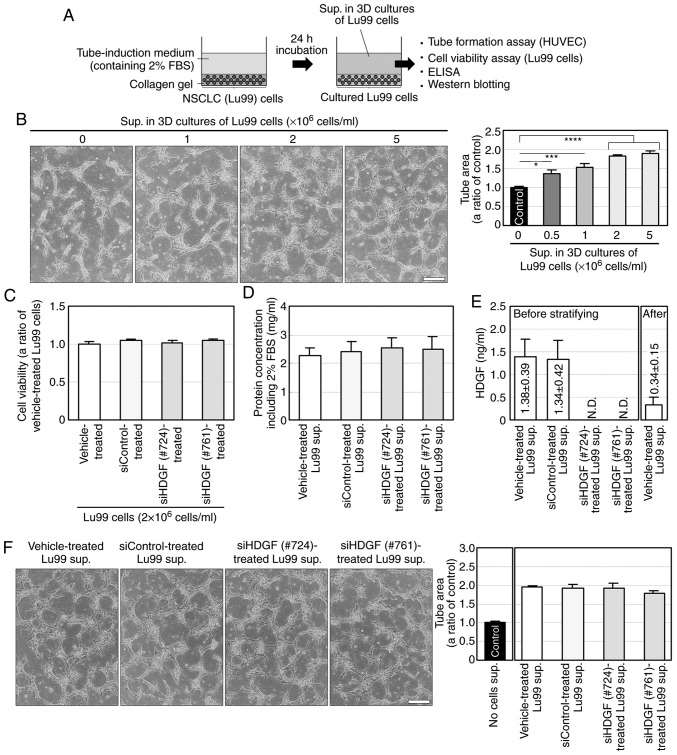 Fig. 3 Lu99 supernatant (sup.) induces HDGF-independent tube formation. (Eguchi R, et al., 2020)
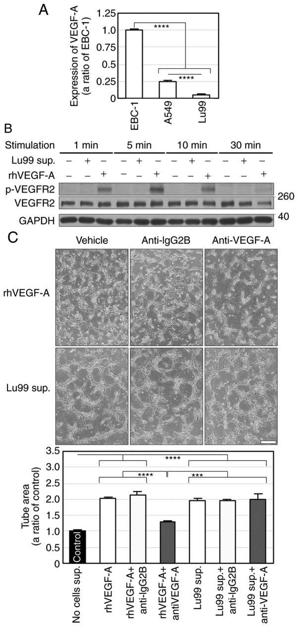 Fig. 4 Lu99 supernatant (sup.) induces VEGF-independent tube formation. (Eguchi R, et al., 2020)
Cell proliferation is one of the critical factors that regulate development. To develop bodies and organs, cell proliferation of multiple rounds is necessary in all multi-cellular organisms during embryogenesis.
Ask a Question
Average Rating: 4.0 | 1 Scientist has reviewed this product
High-quality
As a cancer biologist, I found the tumor cell product I purchased from Creative Bioarray to be of excellent quality.
10 Jan 2023
Ease of use
After sales services
Value for money
Write your own review
- You May Also Need

