Featured Products
Our Promise to You
Guaranteed product quality, expert customer support

ONLINE INQUIRY
LN-405
Cat.No.: CSC-C0317
Species: Human
Source: astrocytoma
Morphology: fibroblastic, grows as monolayers in culture
Culture Properties: monolayer
- Specification
- Background
- Scientific Data
- Q & A
- Customer Review
Immunology: cytokeratin -, cytokeratin-7 -, cytokeratin-8 -, cytokeratin-17 -, cytokeratin-18 -, cytokeratin-19 -, desmin -, endothel -, EpCAM -, GFAP -, neurofilament -,
Glioblastoma multiforme (GBM) is one of the most prevalent primary central nervous system malignancies in adults, with a high proliferative rate, aggressive nature, resistance to radiation and chemotherapy, and limited patient survival. GBM cells have the ability to breach physical walls, including the basement membrane, extracellular matrix and cell junctions, making them one of the most vascularised and aggressive cancers in the human body.
LN-405 is a glioblastoma cell line isolated from a 62-year-old astrocytoma patient. It is the most pervasive glioblastoma type, a WHO grade IV astrocytoma, and one of the most common gliomas in humans. It produces the TGF-1 and G-TSF/TGF-2 mRNA, but not the TGF-1 polypeptide. In GBM, transforming growth factor (TGF)- drives the malignant character by increasing invasiveness, angiogenesis and immunosuppressive microenvironment. Additionally, LN-405 also expresses glial fibrillary acidic protein (GFAP), a principal intermediate filament protein of glia involved in the remodeling of the cytoskeleton and strongly associated with GBM progression.
Since it is extremely aggressive, LN-405 is a prime candidate for understanding how GBM invasiveness works. In vitro studies enable investigators to model the tumour microenvironment, study the biology of tumour cells, and contribute theoretical and experimental evidence to clinical treatment. Additionally, as a model cell line for astrocytoma, LN-405 has wide-ranging applications in pathophysiology, molecular biology and drug development.
Novel Imidazopyridines and Their Biological Evaluation as Potent Anticancer Agents
Güçlü et al. made novel imidazopyridines substituents on both imidazole and pyridine rings, and tested their efficacy against a glioblastoma cell line (LN-405) to determine whether they were glioblastoma targets. Analysing the interactions of the compounds with cells revealed that the most effective molecules were 5a and 5i, both cytotoxic against the LN-405 cell line (Fig. 1A). Flow cytometry was then used to measure the 5a and 5i effects on the cell cycle. The results indicated that the percentage of LN-405 cells in the G0/G1 phase was almost the same for both compounds. While a very low percentage of the cells incubated with 75 µM of 5i were able to reach the S phase, none of the cells incubated with 10 µM of 5a were detected in the S phase. This suggests that 5a and 5i effectively inhibited DNA replication (Fig. 1B). Flow cytometry results indicated that the lead compounds 5a and 5i might have antiproliferative effects on the LN-405 cell line.
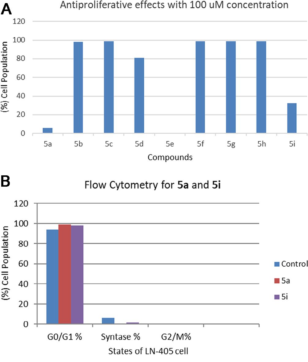 Fig. 1. (A) Cell viability after incubating LN-405 cell lines with 100 µM concentrations of compounds 5a–i. (B) Flow cytometry results for 5a and 5i (Güçlü, D., Kuzu, B., et al., 2018).
The Combination of SB and CEL Affects DNA Repair in Glioblastoma Cell Lines
Sodium butyrate (SB) is a histone deacetylase inhibitor and celastrol (CEL) is an mTOR/protease inhibitor, derived from the tripterygium wilfordii plant. Kartal et al. discovered that SB and CEL inhibit glioblastoma cancer cells by inhibiting DNA repair, apoptotic suppression, and autophagy. On the glioblastoma cell lines LN-405 and T98G, the DNA repair genes MGMT, MLH-1, MSH-2 and MSH-6 were evaluated for their levels. The results indicated significant differences between the control group and the SB, CEL, and SB+CEL cell lines in MGMT, MLH-1, MSH-2, and MSH-6 gene expression (Fig. 2). Additionally, MGMT and MLH-1 gene expression differed between the CEL group and the SB and SB+CEL groups in the T98G cell line (Fig. 2A and B). On the LN-405 cell line, there was a clear difference in MLH-1 expression between the SB+CEL group and the SB and CEL groups (Fig. 2B). Additionally, they observed significant differences in the expression of MSH-2 and MSH-6 in the SB+CEL group versus both the SB and CEL groups in both LN-405 and T98G cell lines (Fig. 2C and D).
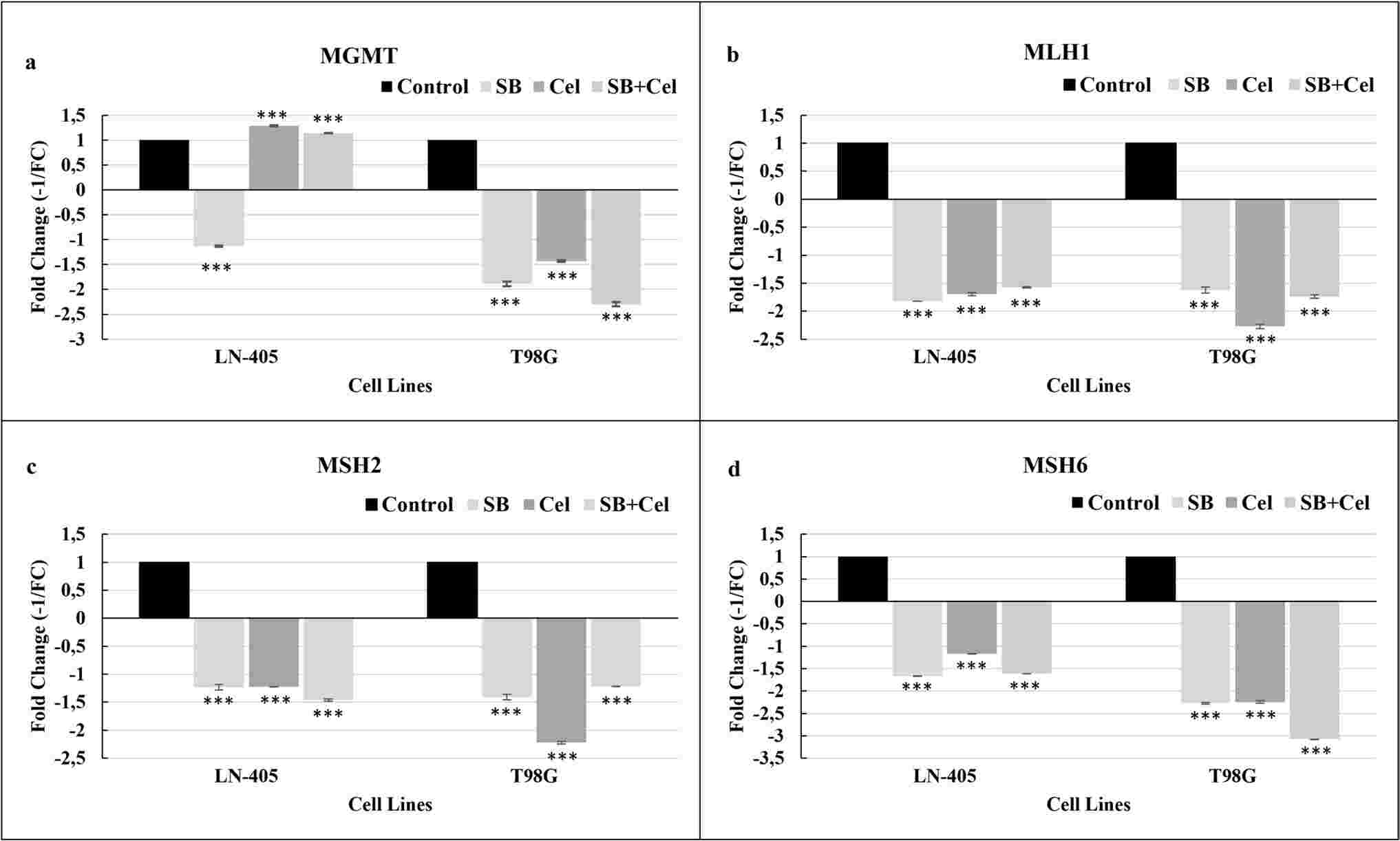 Fig. 2. DNA repair gene expression levels in groups treated with SB, CEL, and SB+CEL combination in LN-405 and T98G cell lines (Kartal B, Denizler-Ebiri FN, et al., 2024).
Open the cap of the bottle in the ultra-clean bench and wipe the outside of the mouth of the bottle with a cotton ball soaked in 75% alcohol to prevent contamination of the cells with spilled medium.
Ask a Question
Average Rating: 5.0 | 1 Scientist has reviewed this product
Perfect
These cells are of high quality and allow reproducible results up to numerous passages.
02 Dec 2020
Ease of use
After sales services
Value for money
Write your own review
- You May Also Need

