CW-2
Cat.No.: CSC-C9175W
Species: Homo sapiens (Human)
Source: Intestine; Colon
Morphology: epithelial-like
- Specification
- Background
- Scientific Data
- Q & A
- Customer Review
The CW-2 cell line is derived from colon carcinoma and serves as an important model for studying colorectal cancer biology. A distinguishing feature of the CW-2 line is its positivity for carcinoembryonic antigen (CEA), a widely recognized tumor marker often associated with colorectal cancers. The presence of CEA indicates that the CW-2 cells can express characteristics typical of malignant colorectal epithelial cells, making them a relevant tool for investigating the mechanisms underlying cancer progression, response to therapies, and the potential for metastasis.
This cell line is also notable for its ability to be transplanted into nude mice, which are immunocompromised and lack a functional immune response. This feature allows for the study of tumor growth and behavior in a living organism, providing insights into the interactions between cancer cells and the host microenvironment. The use of nude mouse models is essential in assessing the efficacy of new therapeutic agents, exploring tumor-stroma interactions, and understanding the metastatic potential of gastrointestinal tumors.
CW-2 Cell Growth and Morphologies in the Culture Containing Aspirin
Cell growth and morphology, which are indexes indicating cell behavior and function, were investigated by phase-contrast micrographies of CW2 cells. Fig. 1 shows the micrographs of CW2 cells on a culture dish made of polystyrene 24 hr after inoculation in the medium containing 0-7.5 mM aspirin at 37°C using 2.0x10⁴ cells/cm2 initially. The cell density decreased with the increase in concentration of aspirin in the medium, and the cells exhibited more spherical morphology in the medium containing a higher concentration of aspirin. The growth kinetics of CW2 cells cultured in the medium containing 0-7.5 mM aspirin were investigated. The growth kinetics are shown in Fig. 2. The cell density was found to decrease with the increase in concentration of aspirin in the medium at any incubation time investigated. In Fig. 2, the cell density increases up to 240 hrs and shows approximately constant cell density after 240 hrs of inoculation, when the cells are cultured in the medium containing less than 3 mM aspirin. On the other hand, cell density remains constant or decreases up to 200 hrs and is constant after 200 hrs of inoculation for the cells cultured in the medium containing more than 3 mM aspirin. LD50, the chemical concentration allowing 50% cell survival, of aspirin is estimated to be 2.0 ± 0.5 mM from a dose-response plot for aspirin based on the cell density after inoculation for 168 hr in Fig. 2.
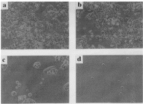 Fig. 1 Micrographs of CW2 cells on culture dishes made of polystyrene after 24 hours of cultivation in RPMI 1640 medium containing 10% FBS and 0 mM (a), 2 mM (b), 5 mM (c), and 7.5 mM (d) aspirin. (Higuchi A, et al., 1999)
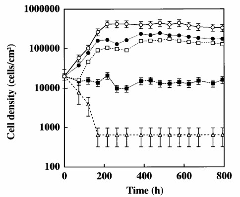 Fig. 2 Growth curves of CW2 cells on culture dishes made of polystyrene in RPMI 1640 medium containing 10% FBS and 0 mM, 2 mM, 3 mM, 5 mM, and 7.5 mM aspirin. (Higuchi A, et al., 1999)
Downregulation of GnT-V Reduces the Sensitivity of CW-2 Cells to Oxaliplatin
GnT-V expression was stably knocked down in the CW-2 and oxaliplatin-resistant CW-2/R cell lines using shRNA. Successful knockdown was verified by western blotting, which showed decreased GnT-V levels in the cells transfected with shRNA#1 and 2 as compared with the wild-type and negative control (NC) cells. Correspondingly, reduced GnT-V activity in the shRNA#1 and 2 cells compared with the NC cells was confirmed by oligosaccharide analyses in cells stained with PHA-L, a lectin that specifically binds to surface β-1,6-N-acetylglucosamine (GlcNAc) branches (Fig. 3A, B). To evaluate the role of GnT-V in chemosensitivity, NC and shRNA#2 transfected cells were exposed to different concentrations of oxaliplatin for 48 h, and then cell viability was determined by CCK-8 assay. As shown in Fig. 3C, the knockdown of GnT-V increased the viability of oxaliplatin-treated CW-2 cells, even at the highest concentration of oxaliplatin (16 µg/ml) compared with the NC group, and a similar result was observed in the CW-2/R group. Moreover, a colony formation assay was performed (Fig. 3D, E). Notably, oxaliplatin-treated shRNA#2 transfected cells exhibited increased survival and clonogenic potential compared with the NC controls. These results suggest that GnT-V knockdown attenuates the chemosensitivity of cells to oxaliplatin.
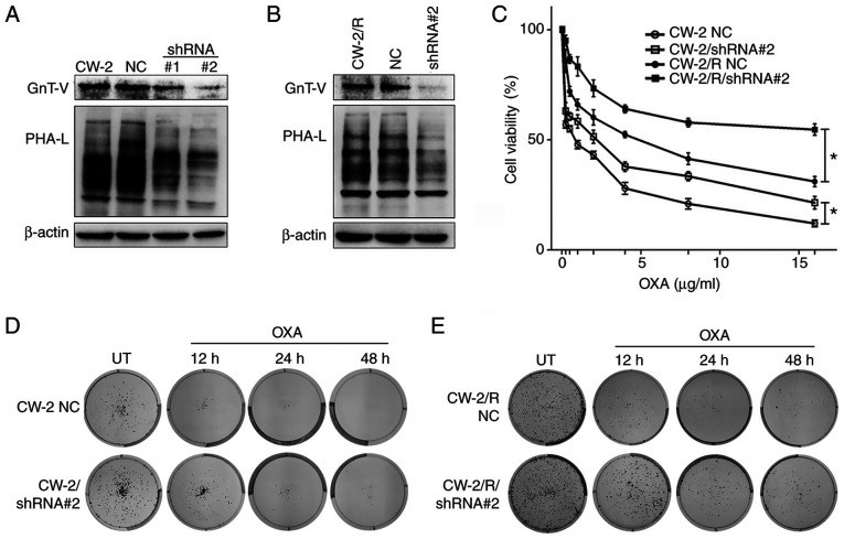 Fig. 3 Cells with GnT-V knockdown exhibit enhanced survival and cell viability upon exposure to oxaliplatin. (A) and (B) shRNA mediated GnT-V knockdown and β-1,6-oligosaccharide reduction in (A) CW-2 and (B) CW-2/R cells as depicted by western blotting and lectin blotting, respectively, compared with the respective parental cell lines and NC cells. (C) Cells were exposed to indicated concentrations of oxaliplatin (0.25-16 µg/ml) for 48 h, and cell viabilities were determined by Cell Counting Kit-8 assay. Representative images of (D) wild-type and (E) drug-resistant cells showing that oxaliplatin-treated GnT-V knockdown cells had reduced chemosensitivity, resulting in an increased colony-forming potential compared with NC cells. (Cong X, et al., 2021)
Ask a Question
Write your own review
- You May Also Need
- Adipose Tissue-Derived Stem Cells
- Human Neurons
- Mouse Probe
- Whole Chromosome Painting Probes
- Hepatic Cells
- Renal Cells
- In Vitro ADME Kits
- Tissue Microarray
- Tissue Blocks
- Tissue Sections
- FFPE Cell Pellet
- Probe
- Centromere Probes
- Telomere Probes
- Satellite Enumeration Probes
- Subtelomere Specific Probes
- Bacterial Probes
- ISH/FISH Probes
- Exosome Isolation Kit
- Human Adult Stem Cells
- Mouse Stem Cells
- iPSCs
- Mouse Embryonic Stem Cells
- iPSC Differentiation Kits
- Mesenchymal Stem Cells
- Immortalized Human Cells
- Immortalized Murine Cells
- Cell Immortalization Kit
- Adipose Cells
- Cardiac Cells
- Dermal Cells
- Epidermal Cells
- Peripheral Blood Mononuclear Cells
- Umbilical Cord Cells
- Monkey Primary Cells
- Mouse Primary Cells
- Breast Tumor Cells
- Colorectal Tumor Cells
- Esophageal Tumor Cells
- Lung Tumor Cells
- Leukemia/Lymphoma/Myeloma Cells
- Ovarian Tumor Cells
- Pancreatic Tumor Cells
- Mouse Tumor Cells
