Featured Products
Our Promise to You
Guaranteed product quality, expert customer support

ONLINE INQUIRY
COLO-678
Cat.No.: CSC-L0003
Species: Human
Source: colon carcinoma
Morphology: round or spindle-shaped adherent cells growing as monolayer
Culture Properties: monolayer
- Specification
- Background
- Scientific Data
- Q & A
- Customer Review
Immunology: cytokeratin +, cytokeratin-7 -, cytokeratin-8 +, cytokeratin-17 -, cytokeratin-18 +, cytokeratin-19 +, desmin -, endothel -, EpCAM +, GFAP -, neurofilament -,
The COLO-678 cell line is a valuable model system in colorectal cancer research, as it was established from a metastatic lymph node sample obtained from a 69-year-old male patient diagnosed with colon carcinoma. The establishment of this cell line in 1984 provides researchers with a clinically relevant model for studying the molecular characteristics and behavior of metastatic colorectal cancer cells.
Metastatic colorectal cancer is a highly aggressive form of the disease, characterized by the spread of tumor cells to distant sites, such as the lymph nodes, liver, and lungs. The COLO-678 cell line, derived from a metastatic lymph node, offers a unique opportunity to investigate the mechanisms underlying the metastatic process and to explore potential therapeutic strategies targeting this advanced stage of the disease.
The COLO-678 cell line has been widely used in various research applications, including the evaluation of novel therapeutic agents, the exploration of mechanisms underlying drug resistance, and the investigation of the tumor microenvironment's role in supporting metastatic progression. The ability of COLO-678 cells to form tumors when injected into animal models further enhances its utility as a preclinical tool for assessing the in vivo behavior of metastatic colorectal cancer.
ALS Modulates CRC Cell Proliferation and the Active Form of RAS
To determine the effects of Aurora kinases A (AURKA) inhibition on RAS signal output, the effects of alisertib (ALS) on cell viability were examined against a panel of colorectal cancer (CRC) cell lines bearing Kirsten rat sarcoma virus (KRAS) WT, G12D, G12V, G13D, A146T, and BRAF V600E mutations. The KRAS G13D-expressing cell line, HCT116, was the most susceptible to ALS treatment over 48 and 96 h (Fig. 1A and B). Colo-678KRAS G12D, SK-CO-1KRAS G12V, and CCCL-18KRAS A146T were also susceptible but to a lesser extent. KRAS WT and BRAF V600E-expressing cell lines exhibited a moderate response to ALS treatment (Fig. 1A and B). Subsequently, the RAS-GTP level was evaluated following the treatment of cells with ALS. RAF1-RBD was used to pull down the active form of RAS from Caco-2KRAS WT, Colo-678KRAS G12D, SK-CO-1KRAS G12V, HCT116KRAS G13D, CCCL-18KRAS A146T and HT29BRAF V600E cells. In KRAS WT-expressing Caco-2 cells, ALS increased the level of RAS-GTP in a concentration-dependent manner (Fig. 2). In KRAS mutant-expressing cells, ALS increased RAS-GTP level in SK-CO-1KRAS G12V, HCT116KRAS G13D and CCCL-18KRAS A146T cells, whereas ALS decreased the RAS-GTP level in Colo-678KRAS G12D cells. Notably, in HT29 cells expressing BRAF V600E, a constitutively active BRAF mutant commonly found in CRC that does not rely on RAS activation to activate the RAS-RAF-MEK-ERK signal, ALS decreased the level of the active form of RAS. In combination, these results suggested that the inhibition of AURKA exerts different inhibitory effects on cell proliferation and regulates the active form of RAS in a KRAS allele-specific manner.
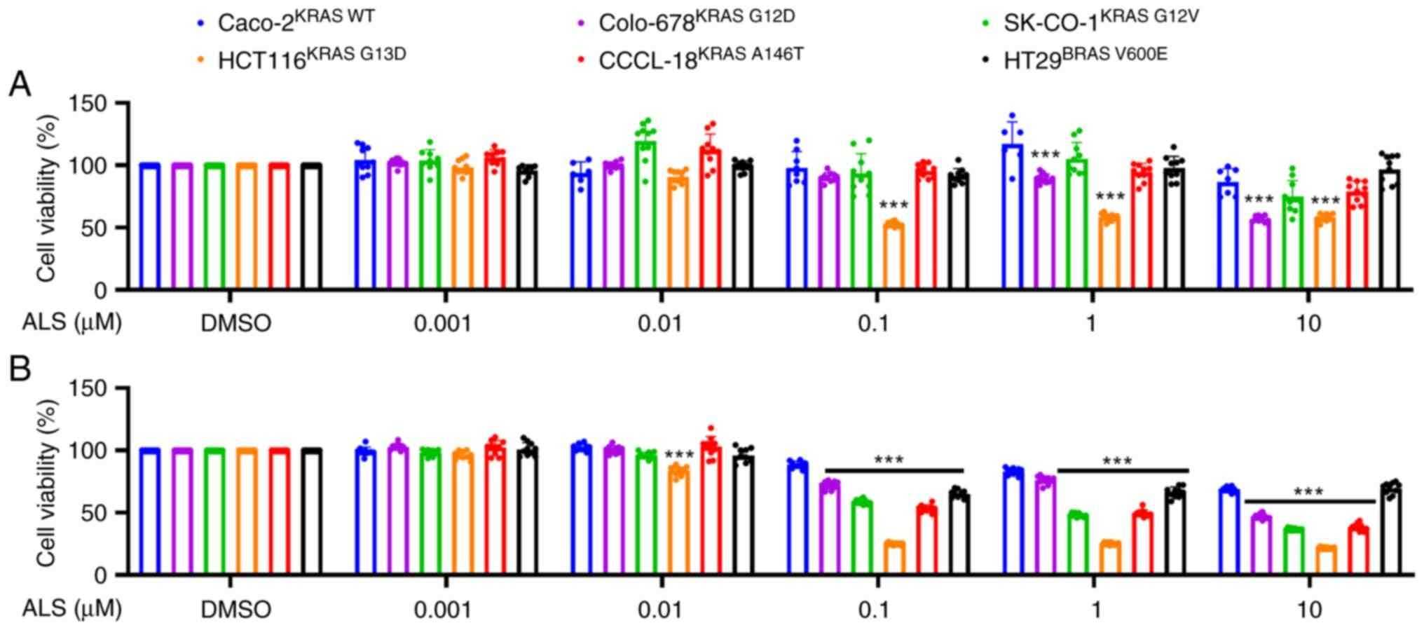 Fig. 1 ALS differentially inhibits the proliferation of CRC cells. (Ren B, et al., 2023)
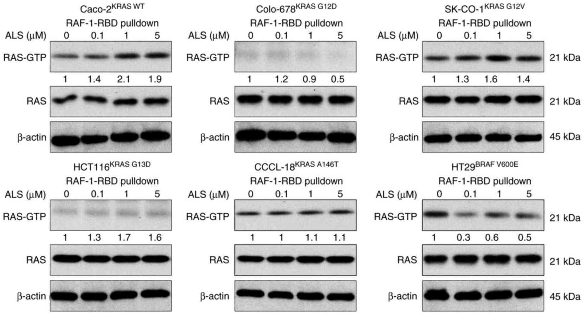 Fig. 2 ALS modulates RAS-GTP level in an RAS allele-specific manner in colorectal cancer cell lines. (Ren B, et al., 2023)
CM from CAFs Exposed to TNF-α Increase Migration of Colo-678 Cells
IL-8 has been previously described to promote the migration of colon cancer cells and endothelial cells in vitro. The increased IL-8 protein levels in cell culture supernatants of cancer-associated stromal fibroblasts (CAFs) exposed to 10 ng/ml TNF-α would increase the migration of these cells in comparison with conditioned medium (CM) of nonstimulated CAFs. As shown in Figure 3, Colo-678 cells showed increased migration toward CM from CAFs incubated with TNF-α in comparison with CM of CAFs that were incubated in 0.5% FBS for the same period. Blockage of IL-8 by neutralizing antibody showed a mild, but not significant, inhibitory effect (Fig. 3C). This lack of evidence was followed for the role of IL-8 as a chemoattractant of the tested cell lines by investigating their migration toward increasing concentrations of IL-8 in the lower chamber. Among the tested cell lines, these experiments revealed only slight increases in migratory activity in Colo-678 cells and HUVECs (data not shown). For the concentration-dependent increases, the lack of responsiveness to IL-8 in our Boyden system strongly indicates that CAFs release other as yet unidentified chemoattractants.
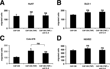 Fig. 3 Effect of TNF-α on chemotactic properties of CAFs. The migration of HuH7 hepatoma cells (A), DLD1 colon carcinoma cells (B), Colo-678 colon carcinoma cells (C), and HUVECs (D) toward the conditioned medium of CAFs that were incubated for 24 hours in 10 ng/ml TNF-α [CAF-CM (TNF)] and toward CM of nonstimulated CAFs (CAF-CM) were compared. (Mueller L, et al., 2007)
Proto-oncogenes are usually genes that help cells grow.
The COLO-678 cell line was established in 1984.
The COLO-678 cell line provides a clinically relevant model for studying the molecular characteristics and behaviors of metastatic colorectal cancer cells, which is crucial for understanding the mechanisms underlying disease progression and developing targeted therapies.
The recommended incubation medium for COLO-678 cells is 90% RPMI-1640 + 10% h.i. FBS.
Ask a Question
Average Rating: 4.7 | 3 Scientist has reviewed this product
Exceed expectation
It exceeded all my expectations and I am so happy I made the purchase.
11 Aug 2022
Ease of use
After sales services
Value for money
Excellent technical support
The team at Creative Bioarray has also been exceptionally responsive and knowledgeable, providing excellent technical support whenever I've had questions or encountered any challenges.
13 Jan 2024
Ease of use
After sales services
Value for money
Reliable suppliers
I have no hesitation in recommending Creative Bioarray as a reliable and trustworthy supplier of high-quality COLO-678 cells.
03 Feb 2024
Ease of use
After sales services
Value for money
Write your own review
- You May Also Need

