Troubleshooting in Fluorescent Staining
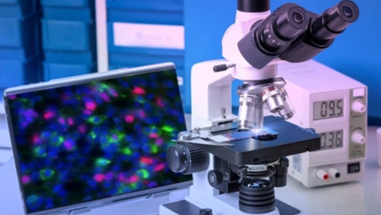
Fluorescent staining plays a pivotal role in life science research, enabling the visualization and analysis of cellular structures and biomolecules with high specificity and sensitivity. However, despite its widespread use, researchers often encounter challenges and issues that can hinder successful imaging.

Troubleshooting Common Issues in Fluorescent Staining
- Insufficient or excessive staining intensity. Achieving optimal staining intensity is crucial for obtaining clear and reliable fluorescence signals. Insufficient staining intensity may result from inadequate antibody concentration, improper incubation conditions, or poor sample preparation. On the other hand, excessive staining intensity can lead to saturated signals, making it challenging to distinguish specific staining from background noise. To troubleshoot these issues, researchers can consider titrating the antibody concentration, optimizing the incubation time and temperature, and ensuring proper sample fixation and permeabilization.
- Non-specific binding and high background fluorescence. Non-specific binding of antibodies and high background fluorescence can arise from various sources, including non-specific antibody interactions, cross-reactivity, or the presence of endogenous fluorophores. To address these issues, researchers can employ blocking reagents to minimize non-specific binding, validate antibody specificity through appropriate controls, and optimize washing steps to remove unbound antibodies or fluorophores thoroughly. Furthermore, careful selection of fluorophores with minimal spectral overlap can help reduce background fluorescence and improve signal-to-noise ratio.
- Uneven or patchy staining pattern. These patterns can arise due to inadequate sample permeabilization, uneven distribution of antibodies, or variations in sample morphology. Researchers can troubleshoot these issues by optimizing the permeabilization step, ensuring thorough mixing of antibodies and samples, and employing gentle agitation during incubation. Additionally, using appropriate controls, such as isotype controls or secondary antibody-only controls, can help differentiate specific staining from non-specific backgrounds.
- Loss of signal during sample processing. This processing can occur due to excessive washing, prolonged exposure to harsh solvents, or inappropriate mounting media. To mitigate this issue, researchers should optimize washing steps to remove unbound antibodies while minimizing signal loss. It is also crucial to handle samples gently during processing and choose mounting media that preserve fluorescence signal integrity. Additionally, proper storage conditions should be maintained to prevent signal degradation over time.
- Autofluorescence and spectral overlap issues. Autofluorescence, resulting from endogenous fluorophores or sample components, can interfere with specific fluorescence signals and lead to false-positive or weak staining. Spectral overlap between fluorophores can also cause signal bleed-through, complicating the interpretation of results. Researchers can address these challenges by selecting fluorophores with minimal spectral overlap, using appropriate controls to distinguish specific signals from autofluorescence, and applying spectral unmixing techniques or advanced imaging technologies to separate overlapping signals.
Troubleshooting Tools and Resources
To assist researchers in troubleshooting fluorescent staining issues, numerous tools and resources are available. Creative Bioarray offers a wide range of validated antibodies, fluorophores, and staining kits, along with comprehensive technical support. These resources guide proper experimental design, troubleshooting tips, and detailed protocols. Additionally, scientific literature, online forums, and collaboration with experienced researchers or core facilities can offer valuable insights and solutions to specific troubleshooting challenges.
Creative Bioarray Relevant Recommendations
Creative Bioarray provides many fluorescent dyes that are highly specific to a variety of organelles and can be used to monitor cell health, cell death, metabolic activity, autophagy, cell tracking, cell migration, and invasion. Our team is always committed to providing customers with high-quality products and services.
| Cat. No. | Product Name |
| FDIR-D0109 | DiA |
| FDIR-D0129 | VitalTracker® ATP Red Live Cell Dye |
| FDIR-D0099 | Calcein AM |
| FDIR-D0133 | Fluo-4 |
| FDIR-D0108 | Nile Red |
| FDIR-D0123 | VitalTracker® Orange Lysosome Dye |
| FDIR-D0102 | VitalTracker® Blue Mitochondria Dye |
| FDIR-D0143 | VitalTracker® Human Pluripotent Stem Cell Dye |
View the details of our fluorescent cellular staining dyes and find what you need!