Protocol for Determining the IC50 of Drugs on Adherent Cells Using MTT Assay
GUIDELINE
Succinate dehydrogenase in the mitochondria of living cells can reduce exogenous MTT to water-insoluble blue-violet crystalline formazan, which is deposited in the cells, but not in dead cells.
Dimethyl sulfoxide (DMSO) can dissolve Formazan in the cells, and its light absorption value was measured at 490 nm with an enzyme marker. Within a certain range of cell number, the amount of MTT crystallization is proportional to the number of living cells, which can indirectly reflect the number of living cells, and quantitatively evaluate the toxicity of the test drug. According to the test data, the semi-inhibitory concentration of the test drug on the cells can be calculated.
METHODS
- Prepare PBS phosphate buffer (1 L, pH = 7.4) containing 0.2 g KCl, 8 g NaCl, 1.44 g Na2HPO4, 0.24 g KH2PO4.
- Weigh 0.5 g of MTT, dissolve in 100 mL of PBS, filter through 0.22 μm microporous membrane, and store at 4°C.
- Approximately 0.5 mg of the drug to be tested was weighed and prepared to an initial concentration of 2.56 mg/mL in DMSO, and the recommended series of gradient concentrations for pre-experimental dilutions were 2.56, 1.28, 0.64, 0.32, 0.16, 0.08, 0.04, and 0.02 mg/mL, respectively.
- Digest the logarithmic phase cells with 0.5% trypsin, when the cells are about to detach from the bottom of the dish, add a small amount of serum to terminate the reaction, centrifuge at 1000 rpm for 5 min, discard the supernatant, and then mix the cell precipitate with medium, count and adjust the concentration of cell suspension to 5-10×104/mL.
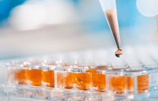
- Add 100 μL of cell suspension into the wells of a 96-well plate, and spread 8 groups of cells per plate, with the concentrations set at 3000, 4000, 5000, 6000, 7000, 8000, 9000, and 10,000 cells/well (the edge of the wells was filled with sterile PBS), and spread a total of 4 plates of cells.
- The 96-well plates were incubated in a 5% CO2 incubator at 37°C, and one plate was taken out every 24 hours for testing. 1) Add 10 μL of MTT solution to each well, and put it back into the incubator to continue incubation for 4-6 hours; 2) Pipette off the culture solution in the wells (the pipette gun should not touch the crystals at the bottom), add 150 μL of DMSO to each well, and shake the plate at low speed for 10 min to fully dissolve the crystals, and then test the absorbance value of the wells at the wavelength of 490 nm with an enzyme counter.
- A standard curve for cell growth was plotted using time as the horizontal coordinate and absorbance values as the vertical coordinate.
- Collect the logarithmic phase cells, adjust the concentration of cell suspension, add 100 μL of cell suspension to the 96-well plate, and spread the plate so that the concentration of cells is 1000-10000 cells/well (the edge wells are filled with sterile PBS).
- Incubate the 96-well plate in a 5% CO2 incubator at 37°C until the cells were attached to the wall, then add a series of drugs with a concentration gradient, the solvent control was DMSO, and the corresponding drugs with known good activity were selected as a positive control, each treatment was repeated 3 times, and the incubation was continued for 24-72 h for microscopic observation.
- Add 20 μL of MTT solution into each well of the 96-well plate, and continue to incubate for 4 h to observe the color development, after terminating the incubation, aspirate the culture solution in the wells, add 150 μL of DMSO into each well, and put it on a shaker for 10 min at a low speed, to make the crystals fully dissolved. The absorbance value of each well was measured at 490 nm by ELISA.
- The semi-inhibitory concentration (SIC) was calculated by the modified Kurtosis method with the formula: 1 g IC50 = Xm - I [P-(3 - Pm - Pn)/ 4] (Xm: 1g maximum dose; I: 1 g (maximum dose/adjacent dose); P: sum of positive response rates; Pm: maximum positive reaction rate; Pn: minimum positive reaction rate).
Creative Bioarray Relevant Recommendations
- Creative Bioarray provides SuperQuick® cell proliferation kit I (MTT) which is a colorimetric assay for the non-radioactive quantification of cellular proliferation, viability, and cytotoxicity. The sample material is either adherent or suspension cells cultured in 96-well microplates.
- We also offer a wide range of cell line testing and assays from cell viability and proliferation to cellular phosphorylation assays. With thousands of cell lines in our biobank and high standard technological platform, most cell assay services have been covered.
NOTES
- MTT is recommended to be used now, and stored at -20°C protected from light after dispensing, if it has turned gray-green, it is not recommended to continue to use.
- To ensure the consistency of the initial density of the plate, the cells need to be counted, according to the counting results, and adjusted to the required density of the plate, the experimental method gives a larger range of different cells that need to be adjusted according to the actual situation, similarly, the range of the starting concentration of the drug need to be pre-experimental judgment.
- The water in the smallest wells at the edge of the 96-well plate evaporates very quickly, which can easily lead to changes in drug concentration and a large error, so it is recommended that the small wells at the edge be filled with sterile PBS.
- At the end of the reaction, the liquid in the wells should be completely discarded. In this method, it is recommended to use a pipette gun to suck up the liquid, or the 96-well plate can be inverted to suck up the liquid with blotting paper, and both methods should be careful not to touch the sediment at the bottom of the wells.