Overview of Cell Apoptosis Assays
Apoptosis (also referred to as programmed cell death) is the event when cells deliberately end their life under certain physiological or pathological circumstances. In apoptosis, cells slowly stop growing and dividing and start killing themselves via the action of some enzymes (like caspases). The hallmarks of apoptotic processes include nuclear split, membrane blebbing, cell fragmentation, the creation of apoptotic masses, and phagocytosis by surrounding cells. Identifying apoptotic activity is vital for disease detection, drug testing, understanding cell death, and keeping tissues in equilibrium. The usual techniques for determining apoptosis are mitochondrial membrane potential measurement and Annexin V/PI staining via biological detection, TUNEL assay and caspase activity measurement via biochemical markers, and morphological observation.
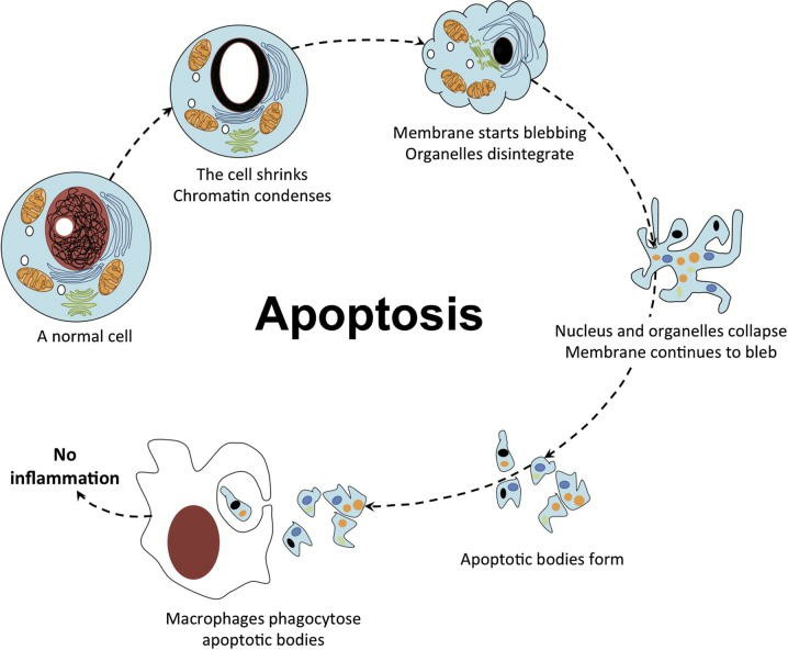 Fig. 1. Cytology of apoptosis (Abou-Ghali M and Stiban J, 2015).
Methods Based on Biological Functions:
- Annexin V/PI Assay: Annexin V is a calcium-dependent phospholipid-binding protein that specifically binds to phosphatidylserine (PS) on the cell membrane. In the early stage of apoptosis, the asymmetry of the cell membrane is disrupted, causing PS to flip from the inner to the outer leaflet of the membrane. Annexin V can bind to this externalized PS. Concurrently, propidium iodide (PI) is a nucleic acid stain that cannot penetrate an intact cell membrane, thus only entering late apoptotic or necrotic cells with compromised membrane integrity, staining the nucleus red. Therefore, the Annexin V/PI dual staining method can distinguish between early apoptotic cells (Annexin V+/PI-), late apoptotic cells (Annexin V+/PI+), and necrotic cells (Annexin V-/PI+).
- JC-1 Assay: In apoptotic cells, increased mitochondrial membrane permeability decreases or eliminates mitochondrial membrane potential (ΔΨm). JC-1 dye detects change in mitochondrial membrane potential; it creates red fluorescent bands at high membrane potential and green fluorescence at low membrane potential.
- Calcium Ion Concentration Assay: This method monitors dynamic changes in intracellular calcium ion concentration during apoptosis using fluorescent calcium ion probes.
- Caspase Activity Detection: The caspase family is crucial in the execution of apoptosis, with their activity significantly increasing during apoptosis. Caspase-3, a key executioner caspase, can be measured using fluorescent probes or ELISA methods to evaluate the extent of cell apoptosis.
Methods Based on Biochemical Markers:
- TUNEL Assay: Apoptosis results in DNA breaks that expose 3'-OH ends. Under the control of terminal deoxynucleotidyl transferase (TdT), these 3'-OH ends attach to a nucleotide labelled with fluorochrome, peroxidase, alkaline phosphatase or biotin.
- Comet Assay: This electrophoretic technique detects DNA migration due to DNA breaks, analyzing DNA damage and fragmentation.
- Bcl-2 Family Protein Detection: The Bcl-2 family of proteins includes pro-apoptotic factors such as Bax, Bak, and Bad, and anti-apoptotic factors such as Bcl-2 and Bcl-xL. Changes in the expression of these proteins can be assessed by Western blot or immunofluorescence to evaluate apoptotic regulation mechanisms.
Methods Based on Morphological Observation:
- Staining Techniques:
Hoechst Staining: Hoechst dyes specifically bind to DNA, resulting in a blue-stained nucleus. In apoptotic cells, nuclear shrinkage and fragmentation occur, altering the stained nuclear morphology, which can be used to observe the morphological characteristics of apoptotic cells.
DAPI Staining: DAPI staining provides clearer visualization of the nucleus and aids in observing nuclear fragmentation and chromatin condensation during apoptosis.
Acridine Orange (AO) Staining: AO staining can differentiate between live and apoptotic cells, with live cells exhibiting green fluorescence and apoptotic cells showing red fluorescence. - Transmission Electron Microscopy (TEM): TEM can be employed to observe organelle changes during apoptosis, such as mitochondrial swelling, endoplasmic reticulum expansion, and nuclear dissolution, which are hallmark features of apoptosis.
- Optical Microscopy Observation: Apoptotic cells typically show reduced cell volume, membrane blebbing, and chromatin condensation and fragmentation, which can be observed via optical microscopy.
- Flow Cytometry: This technique not only detects Annexin V/PI dual staining but also allows for multi-parameter analysis by incorporating other fluorescent markers, such as caspase-3 activity probes, thus providing a more comprehensive evaluation of apoptotic status.
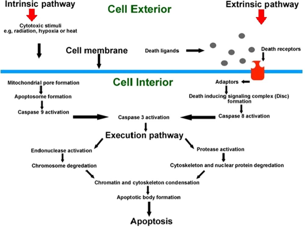 Fig. 2. A summary of the major components of the intrinsic and extrinsic pathways of apoptosis. (D'Arcy MS, 2019).
Creative Bioarray Relevant Recommendations
| Products & Services | Description |
| Cell Apoptosis Assay | Creative Bioarray offers a wide array of apoptosis assays for measuring multiple components on a choice of assay platforms, including Annexin V binding assays, Caspase activity assays, Mitochondrial membrane potential assays, DNA fragmentation and morphology and etc. |
| Cell Apoptosis Assay Kit | Creative Bioarray offers a wide range of assay kits for studying apoptosis including Annexin V Apoptosis Kit, Caspase Assay Kit, DNA Fragmentation Assay Kit, Mitochondrial Apoptosis Detection Kit. |
References
- Asadzadeh Z, Safarzadeh E, et al. Current Approaches for Combination Therapy of Cancer: The Role of Immunogenic Cell Death. Cancers (Basel). 2020. 12(4):1047.
- D'Arcy MS. Cell death: a review of the major forms of apoptosis, necrosis and autophagy. Cell Biol Int. 2019. 43(6):582-592.
- Abou-Ghali M, Stiban J. Regulation of ceramide channel formation and disassembly: Insights on the initiation of apoptosis. Saudi J Biol Sci. 2015. 22(6):760-72.
