Limiting Dilution & Clonal Expansion Protocol
GUIDELINE
To produce a cell line with a particular edit or knockout, clonal expansion of individual cells is necessary. This protocol describes how to isolate single cells from a CRISPR-edited pool, and expand and genotype the subsequent clonal populations.
METHODS
Limiting dilution
- Warm medium at 37°C in a water bath.
- Transfer a 24-well plate containing transfected cells from the humidified CO2 incubator to a biological safety cabinet.
- Aspirate medium from cells across all conditions.
- Carefully wash cells with PBS. Aspirate PBS.
- Add 250 μl cell dissociation reagent to cells.
- Incubate the cells for 5 minutes at 37°C and 5% CO2 or until the cells detach.
- Transfer the well plate back to the biological safety cabinet.
- To neutralize dissociation, add an equal volume of medium (250 μl) to each of the wells.
- Pipet 3-4 times (more if needed) with 1 ml pipette to break up cell clumps. Cells can also be passed through a cell strainer.
- Transfer the cells from each condition to a separate sterile 1.5 ml microcentrifuge tube.
- Count the cells from each condition using a hemocytometer or automated cytometer.
- Calculate the concentration of cells (cells/ml) in the cell suspension.
- Determine the number of 96-well plates for each pool. It is recommended to plate one or two 96-well plates for each edited pool to increase the likelihood that at least one successful knockout clone is generated.
- Dilute cells in a series of 10-fold dilutions to a final concentration of 0.5-1 cells/100 μl of medium (this will give around 1 cell/well in most of the wells in a 96-well plate) or 120 cells in 12 ml of normal growth medium. Scale according to the number of plates desired. For example, if 5 x 96-well plates are desired for a particular condition, dilute 600 cells in 60 ml of normal growth medium.
- Transfer the diluted cell suspension to a large sterile reservoir.
- Dispense 100 μl/well of the diluted cell suspension to each well using a multichannel pipette.
- Repeat steps 13 to 17 for each edited population selected for clonal expansion.
- Transfer plates to a humidified incubator at 37°C, 5% CO2. Change medium as needed.
Clonal expansion
Visually screen plates using a microscope for established single colonies for about two weeks. If a clone image is available, we recommend imaging each of the 96-well plates 2-4 hours after plating the single cell dilution (before the cell can divide). The cells should be imaged 5-7 days thereafter. This practice enables one to track cell growth over time and confirm that the final clones originated from a single cell.
Mark establishing colonies by circling the well on the lid and/or recording it in a spreadsheet. Note that different cells (or possibly genetically modified cells) may grow at a different pace. When colonies reach 70% confluence, transfer them to a 24-well plate. During transfer, take out a portion of cells from each colony for genotyping (see below). By continuing to expand only colonies with confirmed edits, one can reduce the burden of culturing and future screening assays. Maintain a few colonies that grow from the Mock or Negative Control conditions to be used as an isogenic wild-type comparison to the knockout cell line.
Genotyping clones
To genotype your clones, isolate genomic DNA, PCR-amplify the edited region, and sequence the amplicons via Sanger sequencing.
Storage & additional analyses
After identifying putative knockouts by genotype, the cells should be split and expanded for cryopreservation while performing further analysis to functionally validate the knockout. Functional validation of a knockout can be achieved using immunoblot, ELISA flow cytometry analysis, or an appropriate activity assay when available.
Creative Bioarray Relevant Recommendations
- Creative Bioarray provides the world's most comprehensive list of cells and has realized that animal and human primary cells, tumor cell lines, continuous (immortalized) cell lines, and tissues are critical to the biopharmaceutical industry and biomedical research as reagents, therapeutic modalities, and as proxy materials.
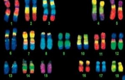
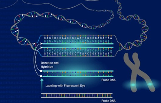
- In recent years proof of clonality has become a focus. Many companies have received comments back on this topic during the IND review process of their biological products. We offer the genetic characterization of producer cell lines by FISH, which offers information on transgene integrity and integration sites.
NOTES
- A clone can be reported as wild type (unedited), containing a homozygous edit, or containing a heterozygous edit. A homozygous edit is when both alleles of a chromosomal pair have the same mutation. These colonies should be saved and marked as potential knockouts. A heterozygous edit is when one allele has a mutation and the other does not (wild type), or when both alleles have different mutations (i.e., compound heterozygous). The heterozygous edit can be easily identified by the overlapping peaks in the chromatogram centered around the region targeted by the guide. Double-stranded breaks repaired by the NHEJ pathway often result in heterozygous edits. This is especially true when editing hyperploid cell lines. Because compound heterozygous mutations may inactivate genes, such colonies should be marked as potential knockouts.
- Whether the edits are of a homo- or heterozygous nature, it is recommended to select colonies containing indels that give rise to frameshift mutations, ideally those that generate a premature stop codon. Indels that maintain the gene's reading frame or alter the coding of only a few amino acids towards the 3' end of the exon, may not lead to the loss of the protein's function.