Resources
-
Cell Services
- Cell Line Authentication
- Cell Surface Marker Validation Service
-
Cell Line Testing and Assays
- Toxicology Assay
- Drug-Resistant Cell Models
- Cell Viability Assays
- Cell Proliferation Assays
- Cell Migration Assays
- Soft Agar Colony Formation Assay Service
- SRB Assay
- Cell Apoptosis Assays
- Cell Cycle Assays
- Cell Angiogenesis Assays
- DNA/RNA Extraction
- Custom Cell & Tissue Lysate Service
- Cellular Phosphorylation Assays
- Stability Testing
- Sterility Testing
- Endotoxin Detection and Removal
- Phagocytosis Assays
- Cell-Based Screening and Profiling Services
- 3D-Based Services
- Custom Cell Services
- Cell-based LNP Evaluation
-
Stem Cell Research
- iPSC Generation
- iPSC Characterization
-
iPSC Differentiation
- Neural Stem Cells Differentiation Service from iPSC
- Astrocyte Differentiation Service from iPSC
- Retinal Pigment Epithelium (RPE) Differentiation Service from iPSC
- Cardiomyocyte Differentiation Service from iPSC
- T Cell, NK Cell Differentiation Service from iPSC
- Hepatocyte Differentiation Service from iPSC
- Beta Cell Differentiation Service from iPSC
- Brain Organoid Differentiation Service from iPSC
- Cardiac Organoid Differentiation Service from iPSC
- Kidney Organoid Differentiation Service from iPSC
- GABAnergic Neuron Differentiation Service from iPSC
- Undifferentiated iPSC Detection
- iPSC Gene Editing
- iPSC Expanding Service
- MSC Services
- Stem Cell Assay Development and Screening
- Cell Immortalization
-
ISH/FISH Services
- In Situ Hybridization (ISH) & RNAscope Service
- Fluorescent In Situ Hybridization
- FISH Probe Design, Synthesis and Testing Service
-
FISH Applications
- Multicolor FISH (M-FISH) Analysis
- Chromosome Analysis of ES and iPS Cells
- RNA FISH in Plant Service
- Mouse Model and PDX Analysis (FISH)
- Cell Transplantation Analysis (FISH)
- In Situ Detection of CAR-T Cells & Oncolytic Viruses
- CAR-T/CAR-NK Target Assessment Service (ISH)
- ImmunoFISH Analysis (FISH+IHC)
- Splice Variant Analysis (FISH)
- Telomere Length Analysis (Q-FISH)
- Telomere Length Analysis (qPCR assay)
- FISH Analysis of Microorganisms
- Neoplasms FISH Analysis
- CARD-FISH for Environmental Microorganisms (FISH)
- FISH Quality Control Services
- QuantiGene Plex Assay
- Circulating Tumor Cell (CTC) FISH
- mtRNA Analysis (FISH)
- In Situ Detection of Chemokines/Cytokines
- In Situ Detection of Virus
- Transgene Mapping (FISH)
- Transgene Mapping (Locus Amplification & Sequencing)
- Stable Cell Line Genetic Stability Testing
- Genetic Stability Testing (Locus Amplification & Sequencing + ddPCR)
- Clonality Analysis Service (FISH)
- Karyotyping (G-banded) Service
- Animal Chromosome Analysis (G-banded) Service
- I-FISH Service
- AAV Biodistribution Analysis (RNA ISH)
- Molecular Karyotyping (aCGH)
- Droplet Digital PCR (ddPCR) Service
- Digital ISH Image Quantification and Statistical Analysis
- SCE (Sister Chromatid Exchange) Analysis
- Biosample Services
- Histology Services
- Exosome Research Services
- In Vitro DMPK Services
-
In Vivo DMPK Services
- Pharmacokinetic and Toxicokinetic
- PK/PD Biomarker Analysis
- Bioavailability and Bioequivalence
- Bioanalytical Package
- Metabolite Profiling and Identification
- In Vivo Toxicity Study
- Mass Balance, Excretion and Expired Air Collection
- Administration Routes and Biofluid Sampling
- Quantitative Tissue Distribution
- Target Tissue Exposure
- In Vivo Blood-Brain-Barrier Assay
- Drug Toxicity Services
Isolation Protocol of Peripheral Blood Mononuclear Cells
GUIDELINE
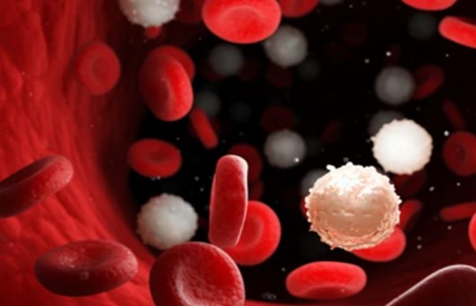
- Peripheral Blood Mononuclear Cell (PBMC), is a cell with a single nucleus in peripheral blood, including lymphocytes, Monocyte, Dendritic Cell (DC), and a small number of other cells. PBMC can be obtained from healthy human or animal donors’ Peripheral blood, obtained by Ficoll density gradient centrifugation (IPHASE/Huiji and source), magnetic bead sorting, and other steps.
- Ficoll is a multimer of sucrose, neutral, with an average molecular weight of 400,000, when the density is 1.2 g/mL, does not exceed the normal physiological osmotic pressure, and does not cross biological membranes. Erythrocytes and granulocytes have a high specific gravity and sink to the bottom of the tube after centrifugation. Lymphocytes and monocytes have a specific gravity less than or equal to the specific gravity of the stratified fluid and float on the liquid surface of the stratified fluid after centrifugation, and a small number of cells may also be suspended in the stratified fluid. Single nucleated cells can be isolated from peripheral blood by aspirating the cells at the surface of the stratified fluid.
METHODS
- Prepare the required solution. The cell culture medium is RPMI 1640 + 10% FBS + 1% P/S and the cell freezing solution is FBS with 10% DMSO.
- Transfer 10 mL of whole blood into a 50 mL centrifuge tube, add 10 mL of PBS solution to dilute, and mix gently.
- Two 15 mL centrifuge tubes are taken and 5 mL Ficoll solution is added first. Then add the diluted blood gently to the upper layer of Ficoll in both centrifuge tubes, making sure to be gentle to avoid mixing the two solutions, 10 mL of diluted blood in each tube.
- Centrifuge at 2,000 rpm for 20 min.
- After centrifugation several layers will be obtained. the cell layer where the PBMC is located is white. At this point, the layer of cells can be pipetted into another clean 15 mL centrifuge tube.
- Add PBS to 10-15 mL, centrifuge at 1500 rpm for 10 min, remove the supernatant, and then add medium for the same wash.
- Add 5-10mL of culture medium to resuspend the cells for subsequent counting culture or plate spreading.
- After collecting the cells by centrifugation, resuspend them with cell lyophilization solution. Take 1-1.5 mL of cells into a lyophilization tube and place it into a lyophilization box (the box can be pre-cooled at 4°C in the refrigerator).
- Place the cassette in the -80°C refrigerator overnight. The next day, transfer the cells to liquid nitrogen for long-term storage.
Creative Bioarray Relevant Recommendations
- Creative Bioarray provides a high-quality and comprehensive range of PBMCs, to accelerate our customers' scientific research, such as Human Normal Peripheral Blood Mononuclear Cells (PBMCs), Human MULTIPLE MYELOMA, Peripheral Blood Mononuclear Cells, Human Peripheral Blood Mononuclear Cells (HPBMC), and others.
NOTES
- The whole blood solution can be added to either the upper or lower Ficoll layer, but ultimately both solutions must be ensured to be delaminated.
- When separating PBMC in the first step of centrifugation, it must not set or set a low level of braking. Otherwise, the stratification will be confusing.
- Asepsis should be observed when drawing human peripheral venous blood.
- The whole operation should be completed in as short a time as possible to avoid increasing the number of dead cells.
- When separating PBMC with lymphocyte separation solution, the increase and decrease of centrifuge speed should be uniform and smooth so that a clear interface is maintained.
- The specific gravity of lymphocytes from mice and rabbits is different from that of humans, so Percoll or different ratios of poly sucrose and pantothenic glucosamine should be prepared with corresponding density.
RELATED PRODUCTS & SERVICES
For research use only. Not for any other purpose.


