Featured Products
Our Promise to You
Guaranteed product quality, expert customer support

ONLINE INQUIRY
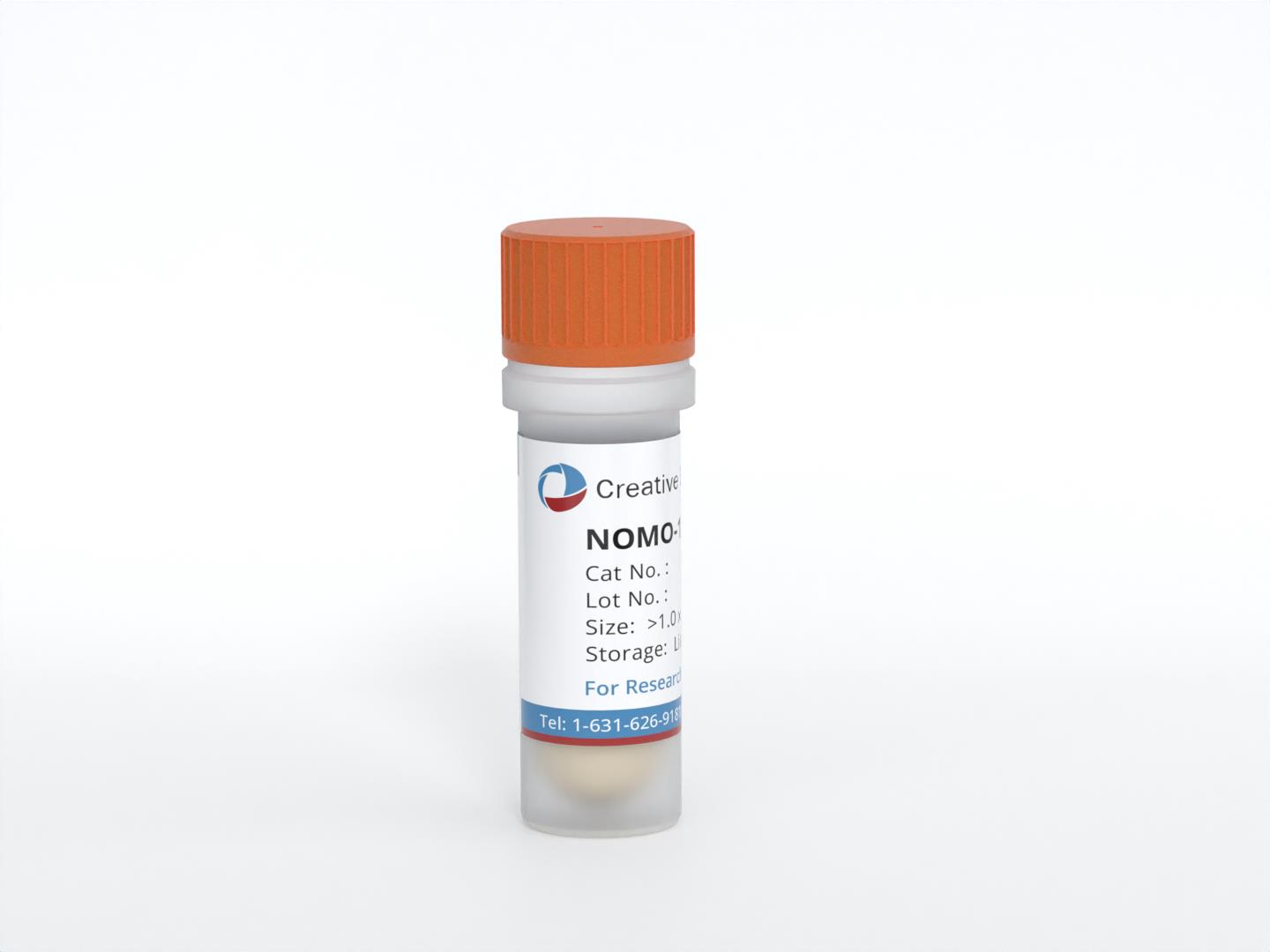
NOMO-1
Cat.No.: CSC-C0550
Species: Human
Source: acute myeloid leukemia
Morphology: single round cells growing in suspension
Culture Properties: suspension
- Specification
- Background
- Scientific Data
- Q & A
- Customer Review
- Documents
Immunology: CD3 -, CD4 +, CD13 +, CD14 -, CD15 +, CD19 -, CD33 +, CD34 -, HLA-DR +
Viruses: PCR: EBV -, HBV -, HCV -, HIV -, HTLV-I/II -, SMRV -
The NOMO-1 cell line is a well-known and extensively studied cell line derived from the bone marrow of a 31-year-old woman with acute myeloid leukemia (AML) at the second relapse. It is classified as AML French-American-British (FAB) subtype M5a, which is characterized by the presence of leukemic cells with monocytic features.
The NOMO-1 cell line exhibits several distinct characteristics that make it a valuable tool in leukemia research. One notable feature is the strong lysozyme activity observed in the cytoplasm of the cells. Lysozyme is an enzyme involved in the degradation of bacterial cell walls and is commonly associated with the phagocytic activity of immune cells. The presence of lysozyme activity in the NOMO-1 cells suggests a monocytic differentiation phenotype.
Additionally, the NOMO-1 cells exhibit phagocytic activities, further indicating their monocytic lineage. Phagocytosis is the process by which cells engulf and internalize foreign particles or microorganisms, contributing to immune defense mechanisms.
NKL Homeobox Gene Activities in NOMO-1 Cell Lines
Frequent deregulation of NKL homeobox genes in myeloid malignancies. For detailed analysis, the NKL homeobox gene NANOG acts as a stem cell factor and is correspondingly expressed alone in hematopoietic progenitor cells.
RQ-PCR and western blot analyses of selected myeloid cell lines confirmed enhanced NANOG expression therein (Fig. 1A). Moreover, the transcript level of stem cell factor NANOG in NOMO-1 matched that quantified in a primary CD34-positive hematopoietic stem cell sample (Fig. 1A), evidencing significant expression of NANOG in this cell line. Therefore, NOMO-1 was chosen as a model for functional analyses of deregulating mechanisms and target genes of NANOG in AML. RQ-PCR analysis of the myeloid NKL-code members DLX2, HHEX, HLX, HMX1, NKX3-1, and VENTX was performed in NOMO-1 in addition to 10 myeloid control cell lines and selected primary cell/tissue samples (Fig. 1B). These data showed that NOMO-1 also expressed elevated HHEX, HLX, and NKX3-1 while the expression of VENTX resembled most controls at a lower level and DLX2 and HMX1 were silenced.
Next, genes potential NANOG-associated candidates were selected and classified these into functional categories including chromatin (BRCA2, EZH2, KMT2A, MLLT3), TFs (BCL6, BHLHE22, DLX5, DLX6, NKX3-1, TP63), NOTCH- and WNT-signalling pathways (CUL1, FBXW5, FZD1, FZD6, HES1, NFX1, NOTCH1), proliferation (CDK6, CDKN2A), and micro-RNA/apoptosis (DROSHA, MIR17HG). RQ-PCR analysis of 10 gene candidates in myeloid cell lines confirmed downregulation of CDKN2A and EZH2 (Fig. 2A) and upregulation of BCL6, CDK6, DLX5, DLX6, FZD1, MIR17HG, NOTCH1 and TP63 in NOMO-1 (Fig. 2B).
 Fig. 1 NKL homeobox gene expression in NOMO-1 cell lines. (Nagel S, et al., 2019)
 Fig. 2 Expression analyses of candidate genes in NOMO-1 cell lines. (Nagel S, et al., 2019)
Pharmacological Inhibition of WIP1 Sensitizes NOMO-1 Cells
In AML, the restoration of p53 activity through MDM2 inhibition proved efficacy in combinatorial therapies. WIP1, encoded from PPM1D, is a negative regulator of p53. The therapeutic efficacy of WIP1 inhibitor (WIP1i) GSK2830371, in association with the MDM2 inhibitor Nutlin-3a (Nut-3a) in AML cell lines and primary samples, was explored.
Single-agent Nut-3a did not reduce cell viability in the TP53-mut cells while showing a time and dosage-dependent effect in all the TP53-wt cells (Fig. 3A), with IC50 values below 1 µM at 72 h (Fig. 3B). WIP1i as a single agent did not significantly affect MOLM-13, HEL, KASUMI-1, and NOMO-1 cell viability (Fig. 3B, C). In line with the cell viability results, apoptosis of NOMO-1, HEL, and KASUMI-1 cells was barely affected by the combined treatment at 48 h (Fig. 4).
The protein level p53 and key signature genes (MDM2 and CDKN1A) were evaluated in the whole panel of cell lines, along with WIP1. In NOMO-1 cells, WIP1 inhibition had mild effects, with no enriched gene sets compared with control cells. Conversely, MDM2 inhibition and the drug combination led to upregulation of inflammatory signatures response (e.g., IL7R and TNFRSF9 upregulated) and TNFA signaling via NFKB (e.g., IER3, PHLDA1 upregulated, Table 1).
 Fig. 3 Viability of AML cell lines treated with Nut-3a and/or WIP1i. (Fontana MC, et al., 2021)
 Fig. 4 Apoptotic response of AML cell line to combined Nut-3a and WIP1i treatment. (Fontana MC, et al., 2021)
Table 1. Most significant enriched pathways in NOMO-1 cells upon drug treatment (FDR: false discovery rate; NES: normalized enrichment score). (Fontana MC, et al., 2021)
| Pathway name | NES | FDR | Treatment Comparison |
| Hallmark of Inflammatory Response | 1.99 | 0.001 | Nut3a+WIP1i vs. DMSO |
| 1.53 | 0.043 | Nut3a vs. DMSO | |
| Hallmark of TNFA Signaling Via NFKB | 2.02 | 0.002 | Nut3a+WIP1i vs. DMSO |
| 1.52 | 0.022 | Nut3a vs. DMSO |
When cells grow old or become damaged, they die, and new cells take their place. Sometimes this orderly process breaks down, and abnormal or damaged cells grow and multiply when they shouldn't. These cells may form tumors, which are lumps of tissue. Tumors can be cancerous or not cancerous (benign).
NOMO-1 cells exhibit strong lysozyme activity in the cytoplasm, display phagocytic activities, and are responsive to differentiation induction with TPA. These characteristics make them a unique cell line for studying AML biology.
NOMO-1 cells carry the t(9;11)(q23;p22) alteration, resulting in the MLL-MLLT3 (MLL-AF9) fusion gene. This genetic anomaly is associated with certain AML subtypes and plays a significant role in leukemogenesis.
NOMO-1 cells can provide valuable insights into AML pathogenesis, cellular responses to differentiation stimuli, and the impact of specific genetic alterations like MLL-MLLT3 fusion gene. Researchers can utilize NOMO-1 cells to study targeted therapies for AML.
Ask a Question
Average Rating: 5.0 | 3 Scientist has reviewed this product
Reasonable ordering
The ordering process was straightforward and hassle-free. The shipping cost was reasonable and justified.
02 Sep 2022
Ease of use
After sales services
Value for money
Knowledgeable and responsive team
The support team of Creative Bioarray was knowledgeable and responsive, making the overall experience extremely positive.
12 Feb 2024
Ease of use
After sales services
Value for money
Preferred supplier
The Creative Bioarray’s professionalism and the quality of its products have made it my preferred supplier for AML research materials.
23 Nov 2023
Ease of use
After sales services
Value for money
Write your own review
- You May Also Need

