Featured Products
Our Promise to You
Guaranteed product quality, expert customer support

ONLINE INQUIRY
HEL
Cat.No.: CSC-C0199
Species: Human
Source: erythroleukemia
Morphology: round, large to occasionally giant polynucleated, single cells in suspension; few cells adherent
Culture Properties: suspension
- Specification
- Background
- Scientific Data
- Q & A
- Customer Review
- Documents
Viruses: ELISA: reverse transcriptase negative;
HEL cells are a human cell line derived from the peripheral blood of a 30-year-old man with erythroleukemia, specifically acute myeloid leukemia subtype M6 (AML M6), in relapse. The patient had previously received treatment for Hodgkin lymphoma. The HEL cell line was established in 1980 and has since been widely used in biomedical research.
These cells are characterized by their ability to undergo spontaneous and induced globin synthesis. This feature makes them valuable for studying the differentiation and maturation of red blood cells, as well as the molecular mechanisms underlying globin gene regulation.
Furthermore, HEL cells are known to carry the JAK2 V617F mutation. This mutation affects the JAK2 gene, which encodes a protein involved in the regulation of cell growth and division. The JAK2 V617F mutation is associated with several myeloproliferative neoplasms, including polycythemia vera and essential thrombocythemia. The presence of this mutation in HEL cells makes them a valuable model for investigating the molecular and cellular aspects of these diseases.
Calothrixin B Derivatives Induce Apoptosis and Cell Cycle Arrest on HEL Cells
Acute erythroleukemia (AEL) is AML characterized by malignant erythroid proliferation. Calothrixin B is a carbazole alkaloid isolated from the cyanobacteria Calothrix and exhibits anti-cancer activity. H-107 has the most potent inhibitory effect on HEL cells, and the IC50 was 3.63 ± 0.33 μM (Table 1). H-107 inhibited HEL cell proliferation in a significant time and dose-dependent manner (Fig. 1A-C). The morphology of HEL cells treated with H-107 gradually shrank and fragmented with the time and concentrations increasing (Fig. 1D). These data suggest that H-107 effectively inhibited HEL cell growth.
Early apoptosis (annexin-V positivity) and late apoptosis (annexin-V/PI positivity) were assessed by flow cytometry. The results displayed that the apoptosis rate of HEL cells treated with H-107 (8 μM) for 48 h was 24.58 ± 0.32%, and the apoptosis rate increased to 32.11 ± 0.68% for 72 h (Fig. 2A and B). Furthermore, H-107 induced HEL cell apoptosis for a significant time and was dose-dependent. The Hoechst 33258 staining indicated that damaged DNA was detected in HEL cells treated with H-107. The result showed that the DNA damage levels were increased in a dose-dependent manner (Fig. 2C).
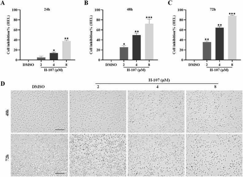 Fig. 1 H-107 inhibited HEL cell proliferation. (Wang B, et al., 2024)
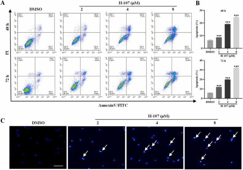 Fig. 2 H-107 induced apoptosis in HEL cells. (Wang B, et al., 2024)
Reactivity of Anti-HEL Cell Monoclonal Antibodies With Normal Hematopoietic Cells
The characteristics of nine monoclonal antibodies (MoAbs) produced using the uninduced cells of a human erythroleukemia line (HEL) as immunogen are described. The proportion of labeled bone marrow cells varied from a very small fraction (antibody 53/10) up to 88% of the cells (antibody 52/40). Based on the frequency of labeled bone marrow cells, the antibodies were classified into four groups, A-D (Fig. 3).
Antibodies of group A (52/10, 52/40, 53/40, 54/ 16) labeled from 43% to 88% of bone marrow cells. In addition, they reacted with large proportions of granulocytes (from 43% to 76%), with 75%-95% of nonadhering non-E rosetting mononuclear cells, and with over 90% of T lymphocytes. The four antibodies failed to react with erythrocytes but did react with erythroblasts. MoAb 53/40 recognized platelets and megakaryocytes.
Antibodies of group B (53/5, 53/6) labeled 17%- 18% of bone marrow cells, a minor population of the non-adhering non-rosetting mononuclear cells, and the monocyte and the T cell fractions, but no granulocytes. The two group B MoAbs failed to react with red cells; they did recognize a small proportion of bone marrow erythroblasts, mainly the immature ones; they labeled strongly the majority of culture-derived erythroblasts.
The two antibodies of group C labeled 100% of the platelets, bone marrow megakaryocytes, and culture-derived megakaryocytic colonies. There was no labeling of red cells or erythroblasts. The antibodies labeled 4%-8% of bone marrow cells and minor populations of granulocytes, monocytes, nonadhering non-rosetting cells, and T cell fractions; whether this small reactivity was due to platelet adsorption on the surface of these cells was not determined.
Antibody 53/10 (the antibody of group D) labeled 1.3% of bone marrow cells, 3.7% of monocytes, 0.8% of non-adhering non-rosetting cells, and 3.7% of T cells. This monoclonal antibody failed to label platelets, erythrocytes, or granulocytes.
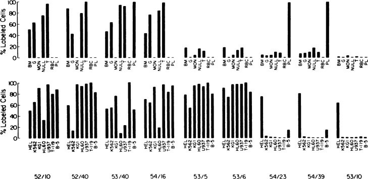 Fig. 3 Reactivity of anti-HEL cell monoclonal antibodies with normal cells (upper panel) and cells of hemopoietic cell lines (lower panel). (Papayannopoulou T, et al., 1984)
Circulating tumor cells (CTCs) are cells that are shed from the primary tumor and circulate in the blood, and their metastasis and formation of a secondary tumor are closely associated with cancer-related death. Therefore, regulating tumor metastasis through CTCs can be a novel strategy to fight cancer.
HEL cells have the ability to undergo both spontaneous and induced globin synthesis. This characteristic makes them useful for studying globin gene regulation and the processes involved in red blood cell development.
HEL cells carry the JAK2 V617F mutation, which is associated with myeloproliferative neoplasms like polycythemia vera and essential thrombocythemia. This mutation affects the JAK2 gene involved in cell growth and division, making HEL cells a valuable model for studying these diseases.
HEL cells provide a valuable model for studying erythroleukemia, allowing researchers to investigate the molecular mechanisms underlying the disease, as well as the differentiation and maturation of red blood cells.
Ask a Question
Average Rating: 4.3 | 3 Scientist has reviewed this product
High-quality
The high quality of Creative Bioarray's cell products ensures consistency in my experiments. I'd highly recommend this to anyone in the field.
05 Apr 2023
Ease of use
After sales services
Value for money
Satisfied
I recently purchased HEL cell products from Creative Bioarray, and I am extremely satisfied with the quality and reliability of their offerings.
01 Jan 2024
Ease of use
After sales services
Value for money
High quality
The cells arrived in excellent condition, maintaining their viability and phenotypic characteristics. I highly recommend Creative Bioarray for its high-quality HEL cell products.
23 Sep 2023
Ease of use
After sales services
Value for money
Write your own review
- You May Also Need

