Featured Products
Our Promise to You
Guaranteed product quality, expert customer support

ONLINE INQUIRY
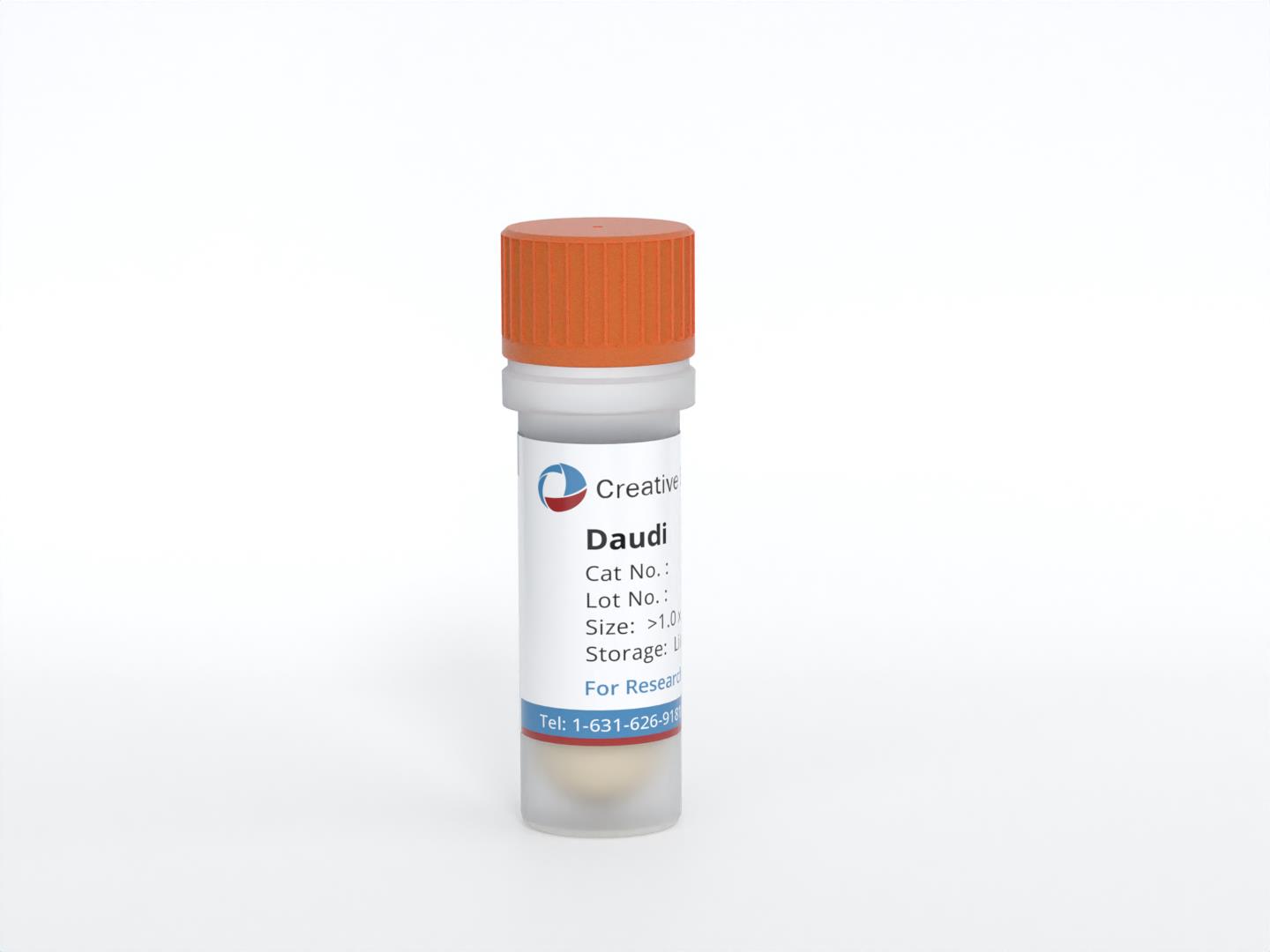
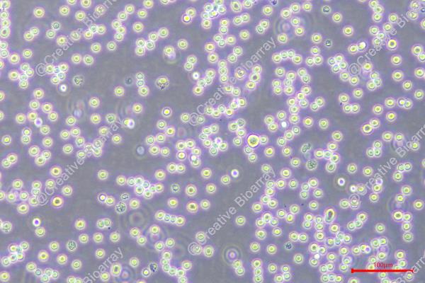
Daudi
Cat.No.: CSC-C9369L
Species: Homo sapiens (human)
Morphology: lymphoblast
Culture Properties: suspension
- Specification
- Background
- Scientific Data
- Q & A
- Customer Review
Receptor: complement; Fc of Ig G
Tumorigenecity: yes, , in nude mice; form colonies in agarose
Isoenzyme: G6PD, B
Karyology: normal male; diploid; stable
Histopathology: lymphoma
Note: Surface immunoglobulin positive. The cells are negative for beta -2- microglobulin and are EBNA positive and VCA. The line carries Epstein-Barr virus
vWA: 15,17
FGA: 21,26
Amelogenin: X,Y
TH01: 6,7
TPOX: 8,11
CSF1P0: 12
D5S818: 8,13
D13S317: 11,12
D7S820: 8,10
The Daudi cell line is composed of B lymphoblasts and was isolated from the peripheral blood of a 16-year-old Black male patient diagnosed with Burkitt's Lymphoma in 1967. This cell line is of particular significance due to its extensive use in the study of leukemogenesis.
The Daudi cells exhibit several unique characteristics that make them a valuable resource for scientific research. They have been confirmed to have tumorigenic properties, capable of forming colonies in agarose and inducing tumors in nude mice. Furthermore, Daudi cells express complement and Fc receptors of Ig G. The cells also possess a normal male, diploid, and stable karyotype. Histopathologically, they are characterized as lymphoma. Interestingly, the Daudi cells are surface immunoglobulin positive, negative for beta-2-microglobulin, and positive for Epstein-Barr nuclear antigens and viral capsid antigens. This suggests that the line carries the Epstein-Barr virus, a factor that could be significant in the study of this type of cancer.
Induction of Apoptosis and CD95 Expression in Daudi Cells by IFN-α
The CD95 receptor, a member of the tumor necrosis factor (TNF) receptor superfamily, mediates signals for cell death on specific ligand or antibody engagement. It was hypothesized that interferon α (IFN-α) induces apoptosis through activation of the CD95-mediated pathway and that CD95 and ligands of the death domain may belong to the group of IFN-stimulated genes.
Evaluation of cell morphology and fluorescence-activated cell sorter (FACS) analysis showed that IFN-α induced cell death in Daudi cells, beginning after 2 days of incubation and increasing to a mean value of 81% (SD ± 0.58%) apoptotic cells after 5 days, compared to a mean value of 11% (SD ± 4.7%) in the control experiment. Apoptosis was preceded by an enhanced expression of CD95 on Daudi cells after 1-day incubation. Receptor expression reached a maximum after 3 days and remained at this level for up to 5 days. Most of the CD95-expressing cells could be detected as apoptotic cells when double staining was applied. The percentage of CD95-TdT double-positive cells rose from a mean value of 14.8% (SD ± 4.7%) after 3 days of IFN-α treatment to a mean value of 83.6% (SD ± 9.3%) after 5 days (Fig. 1).
Because there was a strong correlation between IFN-α-induced apoptosis and CD95 expression, whether IFN-α also stimulates the production and release of CD95L was investigated. However, CD95L was not detected on the cell surfaces of IFN-sensitive Daudi cells by flow cytometry (data not shown). Even after treatment of the cells with a metalloproteinase inhibitor, which is known to block the release of the protein from the cell surface, CD95L was not detectable. In addition, soluble CD95L was not detectable by enzyme-linked immunosorbent assay (ELISA) in supernatants of IFN-treated Daudi cells (data not shown). These results were confirmed by RT-PCR because messenger RNA (mRNA) encoding the CD95L could not be detected in Daudi cells after exposure to IFN-α (Fig. 2).
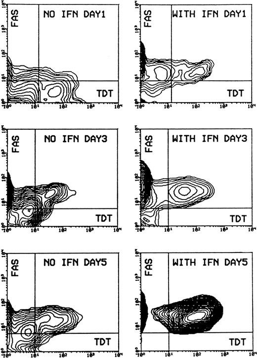 Fig. 1 Induction of apoptosis and expression of CD95 (FAS/APO-1) receptor in Daudi cells in the absence or presence of IFN-α. (Gisslinger H, et al., 2001)
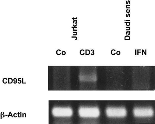 Fig. 2 IFN-α does not induce CD95L mRNA expression. (Gisslinger H, et al., 2001)
The Effect of Anti-CD19 on Daudi Cells
CD19 is a membrane receptor involved in signal transduction in B lymphocytes and it appears on normal B cells early in ontogeny. The effects of different anti-CD19 Abs (HD37, B43.4G7. BU12 [all IgG1]) on the proliferation of Daudi cells in vitro were compared. As shown in Fig. 3, HD37, BU12, and 4G7 inhibited the incorporation of [3H]-thymidinc with an average IC50 of 5.2 ± 2.5 × 10⁻⁷ mol/L(HD37), 5.1 ±0.2 × 10⁻⁷ mol/L (BU12), and 8.9 ± 2.0 × 10⁻⁷ mol/L (4G7). However, although HD37 and BU12 had similar IC50s, BU12 was more effective at killing Daudi cells; at 2 × 10⁻⁶ mol/L BUI2 killed over 90% of the cells, whereas HD37 and 4G7 (at the same concentration) killed only 60%. Therefore, for 4G7 and HD37 to kill as effectively as BU12, threefold more HD37 or 4G7 was necessary (Fig. 3).
B43-anti-CD19, RFB4-anti-CD22, and an isotype-matched control (MOPC-21) did not affect Daudi cells at any concentration tested. The same pattern of inhibition was observed using [3H]-leucine or [3H]-uridine incorporation assays (data not shown). The viability of cells treated with different concentrations of HD37 was also examined. the effect of F(ab')2 and Fab fragments of HD37 on Daudi cells was also compared. The former had the same inhibitory activity as IgG and the latter had no effect at concentrations up to 500 µg/mL (data not shown).
![Daudi cells (1 × 10⁵/well/100 µL) were incubated with different concentrations of Abs (1 × 10⁻⁹ - 1 × 10⁻⁶ mol/L) for 24 hours, then pulsed with [3H]-thymidine for 18 hours, harvested, and counted.](/upload/image/7-daudi-3.jpg) Fig. 3 [3H]-thymidine incorporation in Daudi cells pre-treated with different Abs. (Ghetie MA, et al., 1994)
Fig. 3 [3H]-thymidine incorporation in Daudi cells pre-treated with different Abs. (Ghetie MA, et al., 1994)
Addition of low-temperature organic solvents, low-temperature isotonic solutions, and liquid nitrogen.
Daudi cells have tumorigenic properties, are capable of forming colonies in agarose, and express complement and Fc receptors of Ig G. They are surface immunoglobulin positive, negative for beta-2-microglobulin, and positive for Epstein-Barr nuclear antigens and viral capsid antigens.
Daudi cells possess a normal male, diploid, and stable karyotype.
Histopathologically, Daudi cells are characterized as lymphoma.
Ask a Question
Average Rating: 4.7 | 3 Scientist has reviewed this product
Great
Creative Bioarray's tumor cell products are a great tool for cancer research. Highly recommended!
20 June 2022
Ease of use
After sales services
Value for money
Satisfied
We procured the Daudi cell line from Creative Bioarray for our lymphoma research project, and we have been very satisfied with the quality of the cells.
13 Feb 2024
Ease of use
After sales services
Value for money
Robust growth
The cells have shown robust growth and have met our expectations in terms of performance. I would definitely recommend Creative Bioarray to other researchers.
17 May 2024
Ease of use
After sales services
Value for money
Write your own review
- You May Also Need

