Featured Products
Our Promise to You
Guaranteed product quality, expert customer support

ONLINE INQUIRY
CAL-51
Cat.No.: CSC-C0382
Species: Human
Source: breast carcinoma
Morphology: epithelial-like adherent cells growing in monolayers; the culture contains usually a large amount of cell debris
Culture Properties: monolayer
- Specification
- Background
- Scientific Data
- Publications
- Q & A
- Customer Review
Immunology: cytokeratin +, cytokeratin-7 -, cytokeratin-8 +, cytokeratin-17 -, cytokeratin-18 +, cytokeratin-19 +, desmin -, endothel -, EpCAM +, GFAP -, neurofilament -, vime
Researchers derived the CAL-51 cell line from a pleural effusion metastasis sample taken from a 45-year-old female breast cancer patient whose disease continued to progress after receiving radiotherapy and chemotherapy combined with surgical intervention. The cell line demonstrates epithelial-like properties and adherent growth but certain studies suggest it can also grow in suspension. The cell line exhibits a normal diploid karyotype yet demonstrates microsatellite instability (MSI-low) that results in DNA replication errors which could contribute to tumor development.
Since its establishment in 1985 researchers have used this cell line extensively in breast cancer studies particularly for triple-negative breast cancer research (TNBC). The absence of estrogen receptor (ER), progesterone receptor (PR) and human epidermal growth factor receptor 2 (HER2) expression in this cell line designates it as a subtype of breast cancer with limited treatment choices and poor prognosis. Researchers utilize the CAL-51 cell line to investigate triple-negative breast cancer pathogenesis and biological behaviors like tumor cell proliferation invasion and metastasis and to identify possible therapeutic drugs and treatment plans for this cancer subtype.
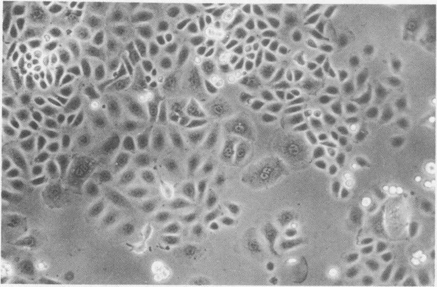 Fig. 1. Photomicrograph of cell line CAL51 (x 70) (Gioanni J, François D L, et al., 1990).
Growth Inhibitory Effect of Everolimus and Gefitinib in TNBC Cell Lines
TNBC shows resistance to both hormonal treatment and anti-HER2 therapy which leads to suboptimal results from standard chemotherapy treatments and early recurrence of disease. The PI3K/AKT/mTOR pathway activation combined with EGFR overexpression occurs frequently in TNBC which presents these molecular targets as potential therapeutic intervention points. Guerrab's team investigated how combining everolimus with gefitinib to block both mTOR and EGFR pathways improves treatment outcomes for TNBC cells, especially those that carry PI3K or PTEN mutations.
The study utilized triple-negative breast cancer cell lines HCC-1937, SUM-1315, and CAL-51 as in vitro models. Everolimus and gefitinib's growth inhibition effects were tested using an XTT assay on three TNBC cell lines, with drug concentrations similar to those in clinical studies. They tested the responsiveness of TNBC cells to ascending everolimus concentrations ranging from 0.1 to 1000 nM as shown in Fig. 1A. At 100 nM concentration cell viability fell by 20% then stabilized with no further decrease even at higher doses while the IC50 remained above 1000 nM across all tested cell lines. The study investigated the combined treatment effects of gefitinib and 100 nM everolimus. Figure 1B demonstrates that cell viability diminished as the dosage increased. The combination of everolimus with gefitinib showed no significant improvement in HCC-1937 and SUM-1315 cells. In CAL-51 cells everolimus enhanced gefitinib's cytotoxic effects significantly across a concentration range of 1 to 20 µM with statistical significance. They examined TNBC cell sensitivity to increasing concentrations (0.1 to 1000 nM) of everolim Comparing experimental results with Bliss theoretical curves showed a synergistic effect in combination treatments. The IC50 of gefitinib alone was 25.15 µM in CAL-51 cells, reduced to 15.49 µM with everolimus.
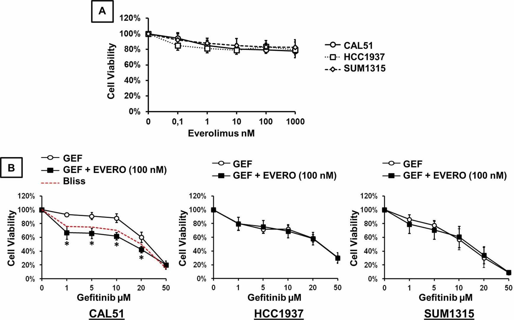 Fig. 1. Cytotoxic effect of gefitinib and everolimus on TNBC cell lines (Guerrab A E, Bamdad M, et al., 2020).
Knockdown of CENPU in TNBC Cells Inhibits Angiogenesis in Vitro
Triple-negative breast cancer (TNBC) is characterized by high vascularity, but anti-angiogenic therapies show poor efficacy. Centromere protein U (CENPU), a centromere component essential for mitosis, is associated with tumorigenesis in multiple cancers; however, little is known of its role in breast cancer. Pan et al. investigated its expression and function of promoting angiogenesis in TNBC.
To investigate CENPU's role in TNBC, they infected MDA-MB-231 and Cal51 cells with lentiviruses carrying CENPU-targeting or control shRNA (shCENPU or CON313) and confirmed successful protein knockdown via western blot (Fig. 2A). RNA sequencing of Cal51-shCENPU and Cal51-CON313 cells showed CENPU's significant association with cancers, especially breast cancer (Fig. 2B). The knockdown of CENPU produced significant effects on genes related to angiogenesis in Cal51 cells according to Gene Ontology analysis (Fig. 2C). The analysis of CENPU's role in angiogenesis and migration included tube formation tests displayed in Fig. 2D and Fig.3B), transwell migration (Fig. 2E, Fig. 3C), and wound-healing assays (Fig. 2F, Fig. 3D). Exposure to TCM from TNBC cells expressing shCENPU led to reduced tube formation and migration abilities as well as wound-healing capacity in HUVECs according to Fig. 2D–F. CENPU-knockdown had limited effects in normal breast cell lines (Figs. 3B–D). This suggests that reducing CENPU expression decreases pro-angiogenic and pro-migratory factors in TNBC cells.
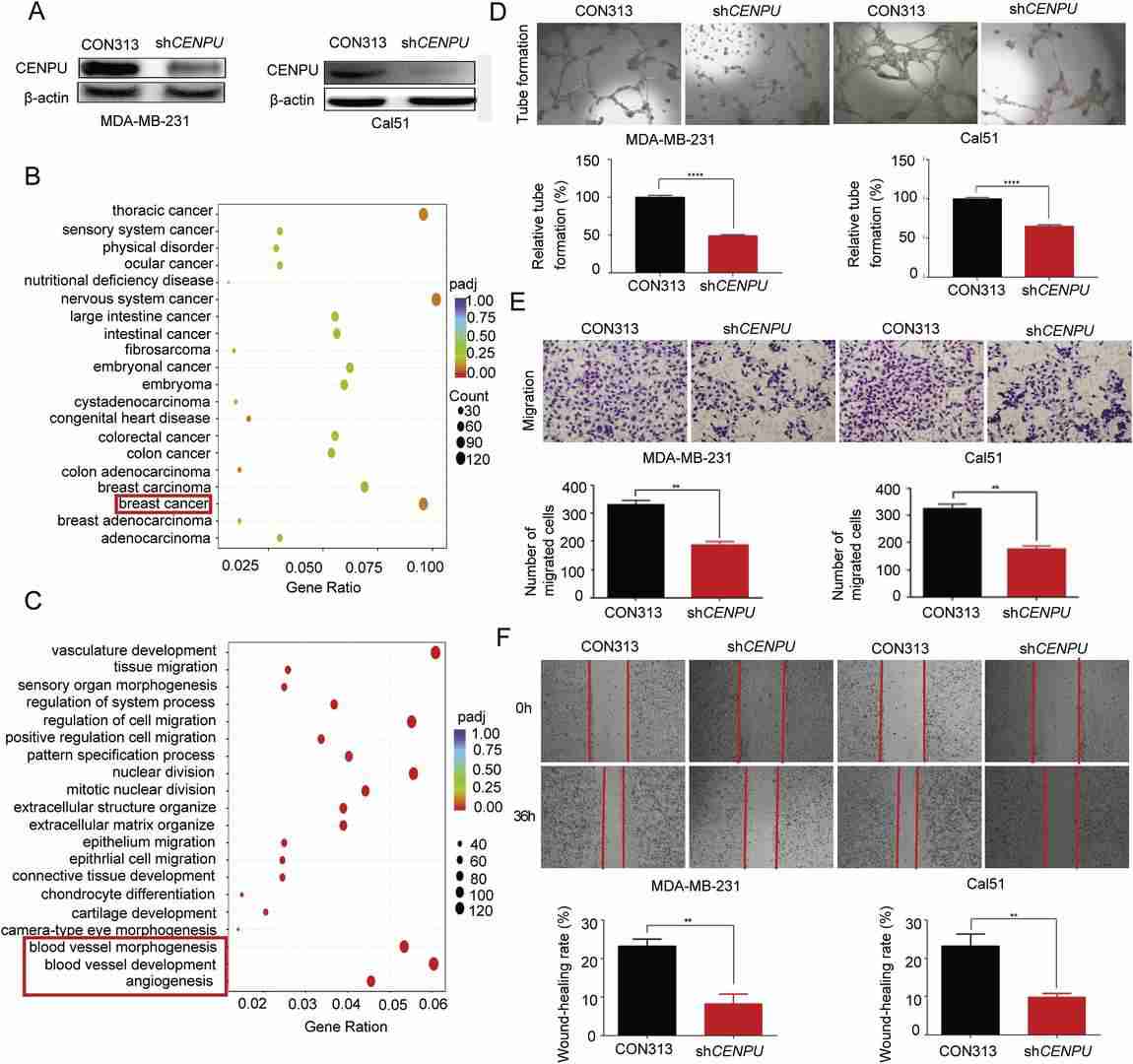 Fig. 2. CENPU promotes TNBC cells angiogenesis (Pan T, Zhou D, et al., 2020).
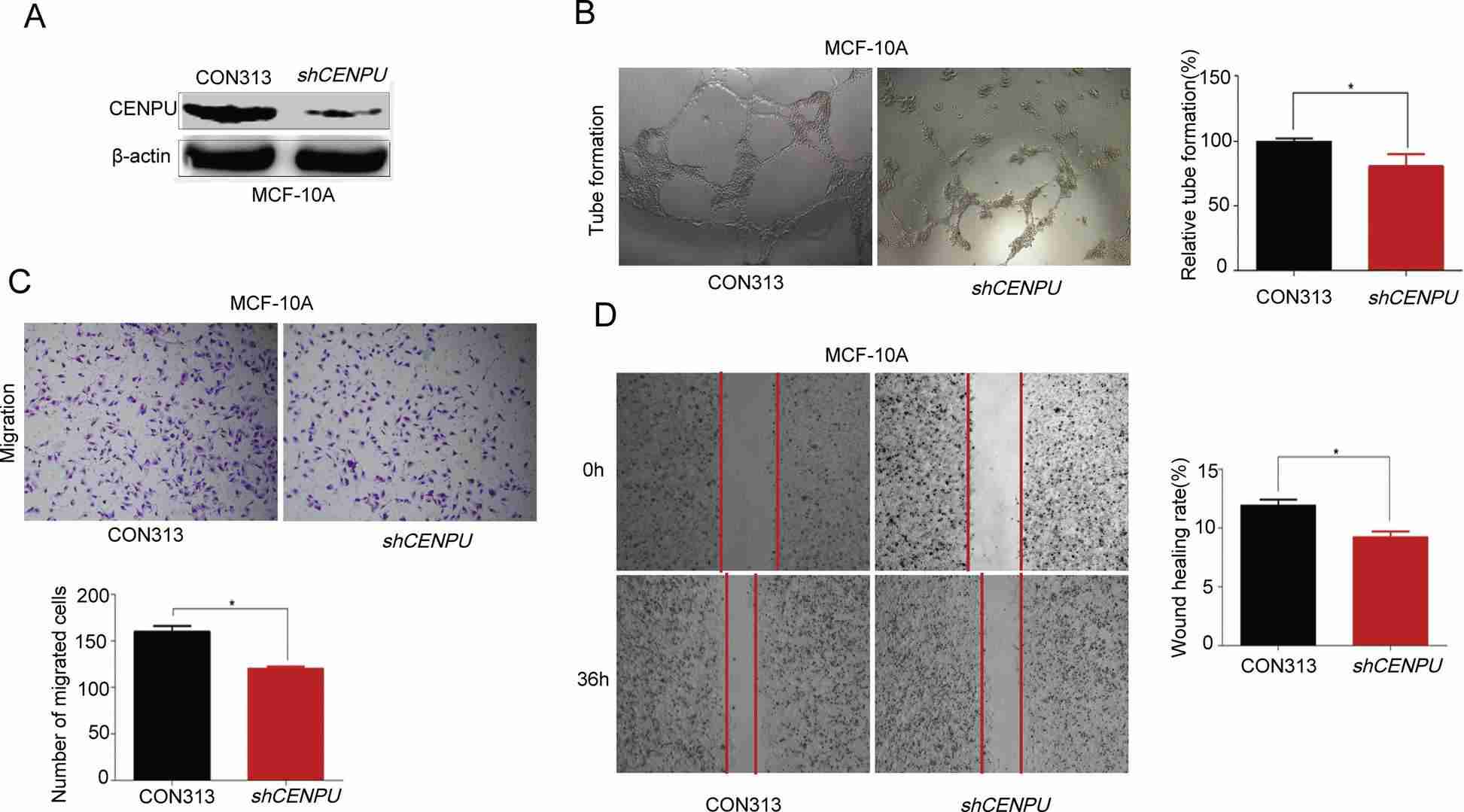 Fig. 3. CENPU promotes normal breast cells angiogenesis limitedly (Pan T, Zhou D, et al., 2020).
The virus may come from the serum.
Ask a Question
Average Rating: 5.0 | 1 Scientist has reviewed this product
Competent platform
Creative Bioarray is a very competent and professional platform.
16 May 2021
Ease of use
After sales services
Value for money
Write your own review
- You May Also Need

Description: Species: human, Caucasian female 69 years old;
Tissue: breast;
Tumor: adenocarcinoma;
Transfected with pBact, expression vector under control of the human beta actin promotor, resistant to G418S.;
Original line: MCF7;
Derived from: pleural effusion

Description: Species: human, Caucasian female 69 years old;
Tissue: breast;
Tumor: adenocarcinoma;
Transfected with ER-pBact, human estrogen receptor cDNA, under control of the human beta actin promotor, resistant to G418S.;
Original line: MCF7;
Derived from: pleural effusion
Description: Species: human, Caucasian female 69 years old;
Tissue: breast;
Tumor: adenocarcinoma;
Transfected with the exon 4 defective variant of ER-pBact cDNA, human estrogen receptor cDNA, under control of the human beta actin promotor resistant to G418S.;
Original line: MCF7;
Derived from: pleural effusion

Description: Species: human, Caucasian female 69 years old;
Tissue: breast;
Tumor: adenocarcinoma;
Transfected with the exon 5 defective variant of human estrogen receptor cDNA, cloned in pZeoSV vector, resistant to zeocin;
Original line: MCF7;
Derived from: pleural effusion

Description: Species: human, Caucasian female 69 years old;
Tissue: breast;
Tumor: adenocarcinoma;
Transfected with the exon 4 defective variant of human estrogen receptor cDNA, cloned in pZeoSV vector, resistant to zeocin;
Original line: MCF7;
Derived from: pleural effusion
Description: Species: human, Caucasian female 69 years old;
Tissue: breast;
Tumor: adenocarcinoma;
Transfected with the pZeoSV-LacZ vector, resistant to zeocin, control for MCF7-488X1 and MCF7-490X1.;
Original line: MCF7;
Derived from: pleural effusion
