
RERF-LC-AI
Cat.No.: CSC-C6515J
Species: Homo sapiens (Human)
Source: Pleural Effusion Metastasis
Morphology: epithelial-like
Culture Properties: Adherent cells
- Specification
- Background
- Scientific Data
- Q & A
- Customer Review
Store in liquid nitrogen.
The RERF-LC-AI cell line is a human lung squamous carcinoma cell line that was established by a patient in Japan. This cell line is part of a broader effort to study lung cancer, particularly squamous cell carcinoma, which is one of the major histological subtypes of non-small cell lung cancer (NSCLC). RERF-LC-AI is notable for its ability to grow in vitro and for its use in various preclinical studies, making it an important model for understanding the biology of lung cancer.
The RERF-LC-AI cell line has been extensively characterized in terms of its genetic and phenotypic properties. The cell line exhibits features typical of squamous carcinoma, including the expression of specific keratin markers and the ability to form distinct cellular structures that resemble the original tumor. These characteristics allow researchers to investigate the molecular mechanisms underlying squamous cell carcinoma development and progression, as well as to explore potential therapeutic targets. In addition to its role in cancer biology, RERF-LC-AI cells are utilized in drug testing and development. Researchers often use this cell line to assess the efficacy of various chemotherapeutic agents and targeted therapies, providing insights into treatment responses and resistance mechanisms.
CLDNs in Human Squamous Carcinoma RERF-LC-AI Cells
Abnormal expression of claudin (CLDN) subtypes has been reported in various solid cancers. The expression levels of CLDN1, 3-5, 7, 12, and 18 were examined in RERF-LC-AI and LK-2 derived from human squamous carcinoma by real-time PCR analysis. In RERF-LC-AI cells, the expression levels of CLDN1 were higher than those in normal tissues, whereas those of CLDN3-5, 7, and 18 were lower (Fig. 1). On the other hand, the expression levels of CLDN7 and 12 in LK-2 cells were higher than those in normal tissues, whereas those of CLDN3, 4, and 18 were lower. RERF-LC-AI cells were used in experiments that followed because the expression pattern of CLDN subtypes in the cells is similar to that in squamous carcinoma tissues.
FLAG-tagged CLDNs were transfected into RERF-LC-AI cells and the expression was determined using anti-FLAG and anti-CLDN3-5, 7, and 18 antibodies. Each CLDN was expressed in the transfected cells (Fig. 2A). The endogenous expression of CLDN1 was not changed by the expression of CLDN3-5, 7, and 18. Immunofluorescence measurements showed that CLDN3 and 4 are mainly distributed in the cytosol and not colocalized with ZO-1 (Fig. 2B). In contrast, CLDN5, 7, and 18 are colocalized with ZO-1, indicating that these CLDNs are distributed in the tight junctions (TJs).
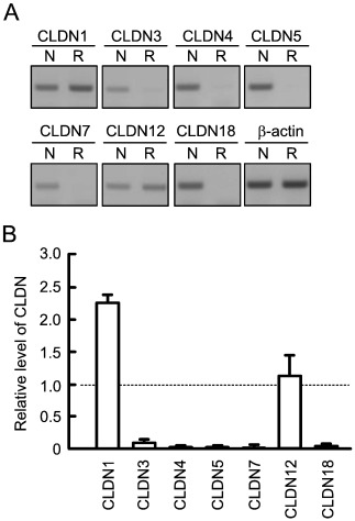 Fig. 1 Expression profile of CLDNs mRNAs in human squamous carcinoma RERF-LC-AI cells. (Akizuki R, et al., 2016)
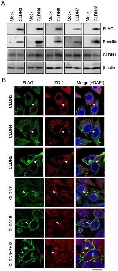 Fig. 2 Expression and intracellular localization of exogenous CLDNs in RERF-LC-AI cells. (Akizuki R, et al., 2016)
Establishment of an Ovarian Metastasis Model Using RERF-LC-AI Cells
The incidence of metastatic ovarian tumors has been reported to comprise 7-10% of all ovarian cancers. The common sources of ovarian metastatic tumors are the stomach, colon and rectum, appendix, breast, uterus, lung, and skin (melanoma). Using transvenous inoculation to NOD/SCID mice, the tumorigenicity of the eight human cancer cell lines (MKN‐28, MKN‐45, MKN‐74, TMK‐1, KATO‐III, HGC27, HSKTC, and RERF-LC-AI) was extremely low in our examination. Almost all cell lines did not show any tumorigenicity, including in the lung or liver. Only RERF-LC-AI, however, formed tumors in several organs (Table 1). RERF-LC-AI metastasized to the lung at 100%, which was considered to be a natural result because this cell line was originally derived from lung cancer, and also because the lung should generally be the first organ that cells reach after injection into the tail vein. RERF-LC-AI most frequently metastasized to the ovary (67%) after the lung, and bilateral lesions were observed in one-third of ovarian metastatic cases (Table 1, Fig. 3a). Therefore, it was considered that RERF-LC-AI had a high propensity for ovarian metastasis and that the experimental system of transvenous inoculation of this cell line to NOD/SCID mice could be used as an in vivo model of ovarian metastasis. Histologically, three-quarters of the ovarian tumors in this model demonstrated a stromal reaction, mimicking the sarcomatoid proliferation of ovarian fibroblasts in the Krukenberg tumor (Fig. 3b).
Ovarian involvement was also observed in these six cell lines; however, in the cases of TMK-1 and MKN-28, the ovaries were buried in the disseminated tumors, and the cancer cells were considered to invade the ovaries directly from predominant adjacently disseminated tumors. On the other hand, by inoculation with HGC27, MKN-45, KATO-III, and RERF-LC-AI, significant enlargement of the ovary without adjacent dissemination was observed (Fig. 3a, c). These four cell lines are considered to have some capacity for metastasizing to the ovary.
Table 1. Organ distribution of experimental metastasis after transvenous inoculation of RERF-LC-AI. (Kuwabara Y, et al., 2008)
| Ovary (unilateral/bilateral) | Peritoneal dissemination | Ascites | Pancreas | Liver | Lung | Bone/bone marrow |
| 3/5 (60%) | 1/5 (20%) | 0/5 (0%) | 1/5 (20%) | 0/5 (0%) | 5/5 (100%) | 1/3 (33%) |
![(a) Macroscopic findings of mouse ovarian tumors. Transevenous inoculation of RERF‐LC‐AI and intraperitoneal inoculation (i.p.) of HGC27 caused bilateral and unilateral enlargement of the ovaries, respectively. In the i.p. experiment of TMK‐1, the ovaries were entirely buried in the disseminated tumor. (b) Histology and immunohistochemistry (smooth muscle actin [SMA] and human-specific cytokeratin) of mouse ovarian tumors of RERF‐LC‐AI. (c) Histology of ovarian tumors established by i.p. of HGC27 and TMK-1.](/upload/image/5-rerf-lc-ai-3.jpg) Fig. 3 Experimental metastasis by inoculation of human carcinoma cell lines. (Kuwabara Y, et al., 2008)
Fig. 3 Experimental metastasis by inoculation of human carcinoma cell lines. (Kuwabara Y, et al., 2008)
Ask a Question
Write your own review
- You May Also Need
- Adipose Tissue-Derived Stem Cells
- Human Neurons
- Mouse Probe
- Whole Chromosome Painting Probes
- Hepatic Cells
- Renal Cells
- In Vitro ADME Kits
- Tissue Microarray
- Tissue Blocks
- Tissue Sections
- FFPE Cell Pellet
- Probe
- Centromere Probes
- Telomere Probes
- Satellite Enumeration Probes
- Subtelomere Specific Probes
- Bacterial Probes
- ISH/FISH Probes
- Exosome Isolation Kit
- Human Adult Stem Cells
- Mouse Stem Cells
- iPSCs
- Mouse Embryonic Stem Cells
- iPSC Differentiation Kits
- Mesenchymal Stem Cells
- Immortalized Human Cells
- Immortalized Murine Cells
- Cell Immortalization Kit
- Adipose Cells
- Cardiac Cells
- Dermal Cells
- Epidermal Cells
- Peripheral Blood Mononuclear Cells
- Umbilical Cord Cells
- Monkey Primary Cells
- Mouse Primary Cells
- Breast Tumor Cells
- Colorectal Tumor Cells
- Esophageal Tumor Cells
- Lung Tumor Cells
- Leukemia/Lymphoma/Myeloma Cells
- Ovarian Tumor Cells
- Pancreatic Tumor Cells
- Mouse Tumor Cells
