ECC10
Cat.No.: CSC-C6443J
Species: Homo sapiens (Human)
Source: Stomach
Morphology: other
Culture Properties: Suspension cells
- Specification
- Background
- Scientific Data
- Q & A
- Customer Review
Store in liquid nitrogen.
The ECC10 cell line is a unique model system derived from a rare and aggressive form of small cell gastrointestinal carcinoma. This subtype of cancer is characterized by the presence of small, undifferentiated tumor cells with a high rate of proliferation and a propensity for early metastasis.
Gastrointestinal small-cell carcinomas are relatively uncommon, accounting for a small percentage of all gastrointestinal malignancies. However, they are often associated with a poor clinical prognosis due to their rapid growth, invasive behavior, and limited treatment options. The availability of the ECC10 cell line provides researchers with a valuable tool to study the underlying mechanisms driving this devastating form of cancer.
A distinguishing feature of the ECC10 cell line is the overexpression of the c-myc oncogene. The c-myc gene is a well-known regulator of cell growth and proliferation, and its dysregulation is frequently observed in a variety of cancers, including gastrointestinal small-cell carcinomas. The overexpression of c-myc in ECC10 cells can contribute to their aggressive phenotype and may serve as a potential therapeutic target.
Interestingly, despite the expected high proliferative capacity associated with c-myc overexpression, the growth rate of the ECC10 cell line is reported to be relatively slow compared to other small cell carcinoma models. This unique characteristic of the ECC10 cells may reflect the complex interplay of various genetic and epigenetic factors that regulate tumor cell growth and behavior.
Ghrelin mRNA Expression in Cultured Cell Lines
To analyze the activity of the human ghrelin promoter, it should be preferable to use a cell line that produces ghrelin endogenously because it seems to contain essential components for the expression of the ghrelin gene. To find a suitable cell line, several cultured cell lines were screened to examine if these cells express ghrelin mRNA endogenously (Fig. 1). In the cell lines examined, TT cell, a human thyroid medullary carcinoma cell line, expressed a high level of ghrelin mRNA endogenously. PCR cycles of 25 times were sufficient for detecting the amplified cDNA fragment of ghrelin. Other cell lines, COS-7, ECC10, HEK293, Hep-G2, and AtT-20, express lower levels of ghrelin mRNA when compared to TT cells. ECC10 and AtT-20 cells need at least 35 cycles of the PCR reaction to detect amplified cDNA fragments of ghrelin. A faint band of the amplified fragment was also observed in HEK293 and Hep-G2 cells after 35 cycles of the PCR reaction. No amplified fragment was observed in the COS-7 cell.
Moreover, the activity of the human ghrelin promoter was higher in TT and ECC10 cells than in COS-7 cells, when the ghrelin promoter/luciferase vectors were transfected into these cell lines (Fig. 2). These results indicate that TT and ECC10 cells that endogenously express ghrelin mRNA contain factor(s), which efficiently activates the human ghrelin promoter.
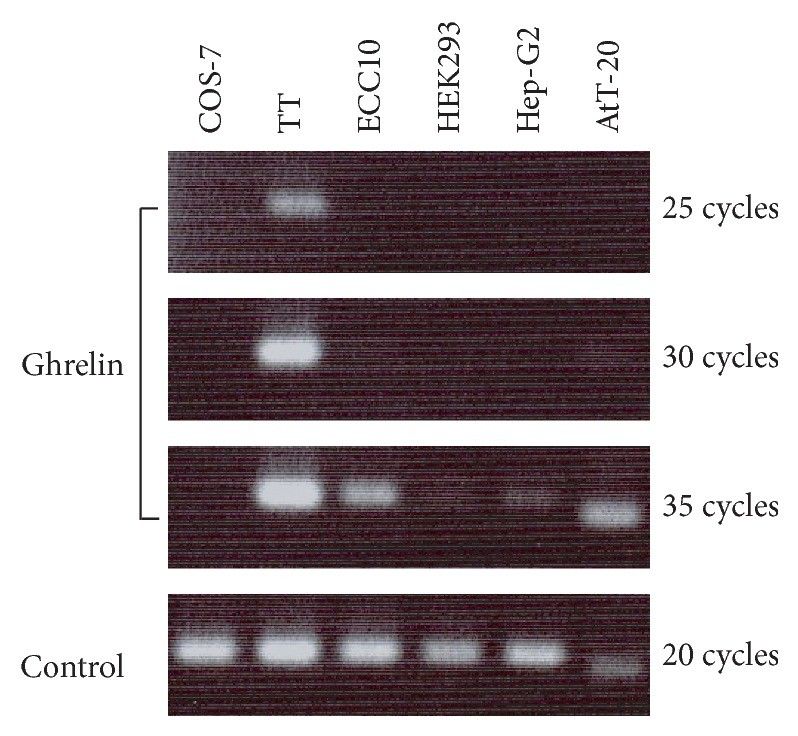 Fig. 1 Expression of ghrelin mRNA in cultured cell lines. A human ghrelin primer set was used for COS-7, TT, ECC10, HEK293, and Hep-G2 cells. A mouse ghrelin primer set was used for the AtT20 cell. Controls are human GAPDH for COS-7, TT, ECC10, HEK293, and Hep-G2 cells and mouse ribosomal RNA18 for AtT-20 cells. (Shiimura Y, et al., 2015)
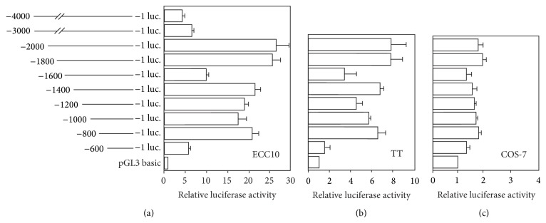 Fig. 2 Functional analysis of human ghrelin promoter in cultured cells. Cell specificity of human ghrelin promoter activity was examined. The human ghrelin promoter sequence with different transcription initiation sites (from -4000/+1 to -600/+1) was ligated to the firefly luciferase and inserted into plasmid pGL3 basic. The constructed vectors were transiently transfected into (a) ECC10, (b) TT, and (c) COS-7 cells, and the luciferase activities were measured. (Shiimura Y, et al., 2015)
MiR-125a-3p Effect on HER2 Protein Expression in Gastric Carcinoma Cells
MiR-125-3p reduces the proliferation and growth of gastric carcinoma cells. Stable transfection data was accomplished (Table 1). The growth rate of cells was significantly reduced in miR-125a-3p transfected gastric carcinoma cells in both cancer cell lines in comparison to other control cells after 48 h, 72 h, and 96 h (maximum inhibition reached after 72 h) of incubation (P < 0.001). In addition, significantly higher growth inhibition percentages were seen in NUGC4 cells compared to the ECC10 cells (P < 0.001). 5-FU treatment together with miR-125a-3p showed a high significance difference from miR-125a-3p alone (P < 0.01) and from other control groups in the same cell line (P < 0.001). The combined treatment of 5-FU with miR-125a-3p showed a significant therapeutic difference in NUGC4 cells in comparison to the ECC10 cells (P < 0.001) (Fig. 3 and 4).
Table 1. Percentages of growth inhibition for cells treated with miR-125a-5p alone or combined with 5-FU (fluorouracil) in comparison to plasmid and RPMI media control groups after 24 h, 48 h, 72 h, and 96 h of exposure in two different gastric cancer cell lines as evidenced by MTT assay. (Mamoori A, et al., 2023)
| Cell line | Treatment | Percent of growth inhibition (Mean±SD) | |||
| After 24 h | After 48 h | After 72 h | After 96 h | ||
| NUGC3 | miR-125a-5p | 15.75±3.16 | 60.09±1.34 | 67.1±1.1 | 68.5±2.34 |
| Plasmid | 12.31±1.69 | 10.6±2.1 | 13.1±1.5 | 9.51±1.2 | |
| S-FU (500 μg/mL) + miR-125a-5p | 66.41±0.67 | 83.06±0.25 | 90.6±1.15 | 89.3±1.0 | |
| Control | 8.47±3.97 | 9.7±1.7 | 11.3±2.2 | 9.9±0.9 | |
| ECC10 | miR-125a-5p | 10.02±1.25 | 37.11±2.7 | 41.56±1.9 | 35.6±2.8 |
| Plasmid | 9.68±3.06 | 14.0±2.1 | 10.7±1.6 | 12.6±1.2 | |
| S-FU (500 μg/mL) + miR-125a-5p | 57.5±0.60 | 64.4±1.15 | 75.2±0.5 | 52.6±1.5 | |
| Control | 11.75±0.70 | 9.32±1.22 | 12.79±2.1 | 10.8±2.2 | |
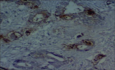 Fig. 3 Moderately differentiated adenocarcinoma of stomach stained slightly for HER2 in IHC analysis. (Mamoori A, et al., 2023)
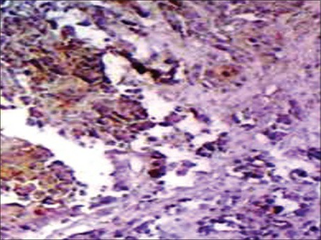 Fig. 4 Poorly differentiated adenocarcinoma of stomach stained strongly for HER2 NEU IHC staining. (Mamoori AMamoori A, et al., 2023)
Ask a Question
Write your own review
- You May Also Need
- Adipose Tissue-Derived Stem Cells
- Human Neurons
- Mouse Probe
- Whole Chromosome Painting Probes
- Hepatic Cells
- Renal Cells
- In Vitro ADME Kits
- Tissue Microarray
- Tissue Blocks
- Tissue Sections
- FFPE Cell Pellet
- Probe
- Centromere Probes
- Telomere Probes
- Satellite Enumeration Probes
- Subtelomere Specific Probes
- Bacterial Probes
- ISH/FISH Probes
- Exosome Isolation Kit
- Human Adult Stem Cells
- Mouse Stem Cells
- iPSCs
- Mouse Embryonic Stem Cells
- iPSC Differentiation Kits
- Mesenchymal Stem Cells
- Immortalized Human Cells
- Immortalized Murine Cells
- Cell Immortalization Kit
- Adipose Cells
- Cardiac Cells
- Dermal Cells
- Epidermal Cells
- Peripheral Blood Mononuclear Cells
- Umbilical Cord Cells
- Monkey Primary Cells
- Mouse Primary Cells
- Breast Tumor Cells
- Colorectal Tumor Cells
- Esophageal Tumor Cells
- Lung Tumor Cells
- Leukemia/Lymphoma/Myeloma Cells
- Ovarian Tumor Cells
- Pancreatic Tumor Cells
- Mouse Tumor Cells
