Featured Products
- HCC-78
- HDLM-2
- DOHH-2
- L-540
- MX-1
- NALM-6
- NB-4
- CAL-51
- SNB-19
- KYSE-520
- MKN-45
- BA/F3
- MS-5
- HCEC-B4G12
- NK-92
- PA-TU-8988S
- MONO-MAC-1
- PA-TU-8902
- Human Microglia
- Human Hepatic Stellate Cells
- Human Skeletal Muscle Cells (DMD)
- Human Schwann Cells
- Human Oral Keratinocytes (HOK)
- Human Cardiomyocytes
- Human Small Intestinal Epithelial Cells
- Human Colonic Epithelial Cells
- Human Intestinal Fibroblasts
- Primary Human Large Intestine Microvascular Endothelial Cells
- Human Small Intestinal Microvascular Endothelial Cells
- Human Retinal Pigment Epithelial Cells
- Human Hepatocytes
- Cynomolgus Monkey Lung Microvascular Endothelial Cells
- Cynomolgus Monkey Vein Endothelial Cells
- C57BL/6 Mouse Primary Mammary Epithelial Cells
- C57BL/6 Mouse Vein Endothelial Cells
- Rat Primary Kidney Epithelial Cells
- Rat Gingival Epithelial Cells
- Rabbit Lung Endothelial Cells
Our Promise to You
Guaranteed product quality, expert customer support

ONLINE INQUIRY

- Specification
- Q & A
- Customer Review
Cat.No.
CSC-C1527
Description
Microglia, one of the glial cell types in the CNS, is an important integral component of neuro-glial cell network. They have been observed in the brain parenchyma from the early stage of development to the mature state. Microglia act as brain macrophages when programmed cell death occurs during brain development or when the CNS is injured or pathologically damaged. Microglia can be considered as the main cell in brain immune surveillance, can present antigens in the molecular context of MHC class II expression to CD-4 positive T cells, are capable of Fc-mediated phagocytosis, and share many common antigens with hemopoietic and tissue macrophages. Furthermore, there is accumulating evidence that microglia are involved in a variety of physiological and pathological processes in the brain by interacting with neurons and other glial cells and through production of biologically active substances such as growth factors, cytokines, and other factors.
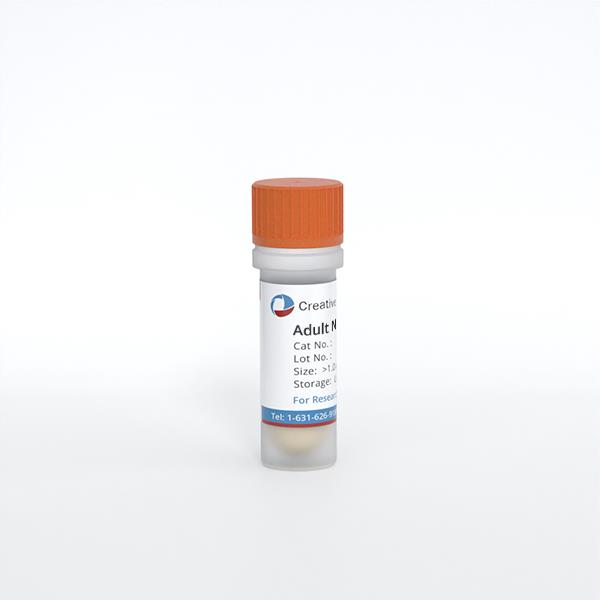
HM are isolated from human brain tissue. After purification, HM are cryopreserved and delivered frozen. Each vial contains >1 x 10^6 cells in 1 ml volume. HM are negative for HIV-1, HBV, HCV, mycoplasma, bacteria, yeast and fungi.
HM are isolated from human brain tissue. After purification, HM are cryopreserved and delivered frozen. Each vial contains >1 x 10^6 cells in 1 ml volume. HM are negative for HIV-1, HBV, HCV, mycoplasma, bacteria, yeast and fungi.
Source
Brain
Recommended Medium
It is recommended to use Microglia Medium for the culturing of HM in vitro.
Cell Type
Microglia
Disease
Normal
Storage and Shipping
ship in dry ice; store in liquid nitrogen
Citation Guidance
If you use this products in your scientific publication, it should be cited in the publication as: Creative Bioarray cat no. If your paper has been published, please click here to submit the PubMed ID of your paper to get a coupon.
Ask a Question
Write your own review
Related Products


