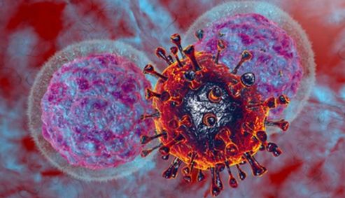
Isolation, Expansion, and Analysis of Natural Killer Cells
Natural killer (NK) cells are a type of lymphocyte that plays a crucial role in the innate immune response against infected and malignant cells. They were first identified in the 1970s and were named "natural killers" due to their ability to spontaneously recognize and eliminate target cells without the need for prior sensitization or activation. These cells are capable of recognizing and eliminating target cells without prior sensitization, making them an important component of immune surveillance. In recent years, there has been significant interest in isolating, expanding, and analyzing NK cells for various therapeutic applications.

What Are NK Cells?
NK cells are a subset of lymphocytes that primarily reside in the peripheral blood, spleen, and lymph nodes. They are characterized by the expression of surface markers such as CD56 and CD16. Unlike T cells, which require antigen presentation and activation, NK cells can directly recognize and eliminate target cells based on a delicate balance between activating and inhibitory signals.
Table 1. Cellular markers expressed during human and mouse NK cell developmental stages.
| Species | Cellular markers |
| Human | CD7, CD10, CD34, CD38, CD56Hi, CD117, CD122, CD127, CD133, CD161, CD244, CD335, CD337, ILR1, NKG2A, NKG2D, etc. |
| Mouse | CD27, CD43, CD117, CD122, CD127, CD244, NK1.1, NKG2D, NKG2A/C, etc. |
NK cells exert their cytotoxic effects through two main mechanisms: direct cell-mediated cytotoxicity and antibody-dependent cellular cytotoxicity (ADCC). In direct cell-mediated cytotoxicity, NK cells recognize cells that display altered surface markers or stress-induced ligands and induce apoptosis in the target cells. In ADCC, NK cells recognize target cells opsonized with antibodies and subsequently trigger the release of cytotoxic granules, leading to target cell lysis.
Source and Isolation of NK Cells
NK cells can be isolated from various sources, including peripheral blood, umbilical cord blood, and tissues such as the spleen and lymph nodes. Peripheral blood is the most commonly used source for NK cell isolation due to its easy accessibility. Several techniques have been developed for the isolation of NK cells, including gradient density centrifugation, magnetic cell sorting, and flow cytometry-based cell sorting.
- Density gradient centrifugation. This technique separates cells based on their density using a density gradient medium. It allows for the separation of PBMCs or dissociated tissue cells from other components, resulting in a fraction enriched in NK cells.
- Magnetic cell sorting. It utilizes magnetic beads conjugated with antibodies specific to NK cell surface markers. The beads bind to the NK cells, and a magnetic field is applied to isolate the bead-bound cells. This technique enables positive selection, negative selection, or depletion of NK cells based on the desired surface markers.
- Flow cytometry-based cell sorting. Flow cytometry can be used to sort and isolate NK cells based on their specific surface marker expression. In this technique, cells are labeled with fluorescently labeled antibodies targeting NK cell markers. The labeled cells are then passed through a flow cytometer, which detects and sorts the cells based on their fluorescence signals.
Culture and Activation of NK Cells
Once isolated, NK cells can be cultured and expanded ex vivo to obtain sufficient cell numbers for various applications. The culture medium used for NK cell expansion typically contains a combination of cytokines, such as interleukin-2 (IL-2), IL-15, and IL-21, which promote NK cell survival and proliferation.
In addition to cytokines, several other factors can influence the expansion and activation of NK cells. Co-culture with feeder cells, such as irradiated PBMCs or artificial antigen-presenting cells (aAPCs), can provide additional stimulatory signals and enhance NK cell expansion. Furthermore, the addition of specific activating agents, such as Toll-like receptor (TLR) ligands or antibodies targeting NK cell receptors, can further enhance NK cell activation and cytotoxicity.
Analysis and Characterization of NK Cells
To fully understand the functional properties of NK cells, it is essential to perform comprehensive analysis and characterization. Flow cytometry is a powerful technique that allows the phenotypic and functional profiling of NK cells. By staining NK cells with a panel of fluorescently labeled antibodies, it is possible to analyze the expression of various surface markers and intracellular molecules.
Phenotypic analysis of NK cells can provide valuable insights into their maturation status, activation state, and functional subsets. For example, the expression of CD56 and CD16 can be used to identify distinct NK cell subsets, such as CD56brightCD16dim and CD56dimCD16bright cells, which exhibit different functional properties.
Functional analysis of NK cells can be performed using various assays, such as cytotoxicity assays, cytokine production assays, and degranulation assays. Cytotoxicity assays measure the ability of NK cells to kill target cells, while cytokine production assays assess the secretion of cytokines, such as interferon-gamma (IFN-γ) and tumor necrosis factor-alpha (TNF-α). Degranulation assays measure the release of cytotoxic granules from NK cells upon target cell recognition.
Table 2. Key cytokines involved in NK cell activation, suppression, and secretion.
| Activation | Suppression | Secreted | |
| Cytokines and chemokines | IL-2, IL-15, IL-12, IL-18, and IL-21 IFG-g | IL-4, IL-5, IL-1β, IL-6, IL-10, IL-23, IL-35, TGF-β | IFN-γ, TNF-α, GM-CSF, IL-10, IL-5, and IL-13, MIP-1α, MIP-1β, IL-8, RANTES |
| Other factors | Granzyme-A, -B, & -M, Perforin |
Creative Bioarray Relevant Recommendations
| Product/Service Types | Description |
| Cell Isolation Kit | Creative Bioarray is dedicated to delivering thoroughly tested and high-quality products that do not adversely affect cells during isolation. |
| Cell Surface Marker Validation Service | Creative Bioarray provides cell surface marker validation services to support all researchers, biotech, and pharmaceutical companies in the progress of drug development. |
| Cell Line Authentication | Creative Bioarray STR profiling is critical for verifying the identity of human cell lines, ensuring the uniqueness of the cell line, and detecting laboratory errors such as misidentification and cross-contamination of lines. The sensitivity and high power of discrimination make our STR analysis an ideal choice for the various types of cell authentication. |
| ActoFactor™ GMP Cytokines | We offer a large variety of GMP-grade cytokines. ActoFactor™ GMP cytokines are manufactured and tested in compliance with relevant GMP guidelines. |